. 2014 Jul;42(12):8024-38.
doi: 10.1093/nar/gku446. Epub 2014 May 23.

Structure and semi-sequence-specific RNA binding of Nrd1

Affiliations

Structure and semi-sequence-specific RNA binding of Nrd1

Veronika Bacikova et al. Nucleic Acids Res. 2014 Jul.

Abstract

In Saccharomyces cerevisiae, the Nrd1-dependent termination and processing pathways play an important role in surveillance and processing of non-coding ribonucleic acids (RNAs). The termination and subsequent processing is dependent on the Nrd1 complex consisting of two RNA-binding proteins Nrd1 and Nab3 and Sen1 helicase. It is established that Nrd1 and Nab3 cooperatively recognize specific termination elements within nascent RNA, GUA[A/G] and UCUU[G], respectively. Interestingly, some transcripts do not require GUA[A/G] motif for transcription termination in vivo and binding in vitro, suggesting the existence of alternative Nrd1-binding motifs. Here we studied the structure and RNA-binding properties of Nrd1 using nuclear magnetic resonance (NMR), fluorescence anisotropy and phenotypic analyses in vivo. We determined the solution structure of a two-domain RNA-binding fragment of Nrd1, formed by an RNA-recognition motif and helix-loop bundle. NMR and fluorescence data show that not only GUA[A/G] but also several other G-rich and AU-rich motifs are able to bind Nrd1 with affinity in a low micromolar range. The broad substrate specificity is achieved by adaptable interaction surfaces of the RNA-recognition motif and helix-loop bundle domains that sandwich the RNA substrates. Our findings have implication for the role of Nrd1 in termination and processing of many non-coding RNAs arising from bidirectional pervasive transcription.


Figures

Figure 1.
Figure 1.
Overview of domain organization of Nrd1, sequence and NMR data of Nrd1307–491. (A) Scheme of the full-length Nrd1 protein containing CTD-interacting domain (CID), dimerization domain (DD), arginine-glutamate/arginine-serine-rich region (RE/RS), RNA-recognition motif (RRM) and proline-glutamine-rich sequence (P/Q). (B) Nrd1307–491 construct and its sequence along with highlighted RNP2 and RNP1 sites and predicted secondary structure elements. (C) 1H-15N HSQC spectrum of Nrd1307–491 measured at 20°C in 50-mM phosphate buffer (pH = 8) supplemented with 300-mM NaCl and 10-mM β-mercaptoethanol. (D) Secondary structure prediction based on Cα and CO chemical shifts correlates with the predicted RRM topology. The plot shows an additional structured region in the C-terminus.
Figure 2.
Figure 2.
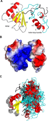
NMR structure of Nrd1307–491. (A) The lowest energy three-dimensional solution structure of Nrd1307–491 consisting of two domains, an RRM with βαββαβ topology and an additional helix–loop bundle domain. The latter domain harbors both N- and C-terminal regions to the RRM of Nrd1 (in black and cyan). The protein is shown as a ribbon model, with β-sheets in yellow and α-helices in red. The structure has been determined using 760 structurally meaningful NOE distance restraints derived from NOESY data acquired on the highly deuterated 2H, 15N, 13C, (Val, Leu, Ile)-methyl, ethyl-protonated protein sample. (B) Solvent-accessible surface representation of the representative structure of Nrd1307–491 colored by electrostatic potential (blue, positive; red, negative). (C) Overlay of the 20 lowest energy structures of the free form of Nrd1307–491 over the RRM domain. Figures were generated with PyMOL (Schrödinger, LLC).
Figure 3.
Figure 3.
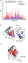
Two types of Nrd1–RNA interaction described by NMR. (A) Comparison of Nrd1307–491 binding to GUAA (red) and G7 (blue) RNA sequences. GUAA RNA is recognized mostly by residues within β-sheet surface, whereas G7 interaction is mediated by amino acids from the additional helix–loop bundle domain. (B) Structure of Nrd1307–491 with highlighted regions that are responsible for AU-rich (red) and G-rich (blue) RNA binding. Overlapping region is shown in magenta. RNA-binding surface was colored based on the mutagenesis results. (C) A canonical RRM binds RNA via its β-sheet surface (red), exemplified here by the structure of sex-lethal RRM1 [PDB code: 1B7F; (55)].
Figure 4.
Figure 4.
In vitro and in vivo mutational study of Nrd1. (A) GUAA binding by the Nrd1307–491 mutants assayed using FA. (B) GCGGGGC binding by the Nrd1307–491 mutants assayed using FA. (C) In vivo phenotypic analyses of the Nrd1 mutants. Wt Nrd1 contains non-mutated NRD1 gene, pRS415 is a negative control with empty plasmid without NRD1 gene and the other plasmids contain NRD1 point mutations as denoted. The indicated mutants were expressed episomally from pRS415 plasmids in the yeast strain with the endogenous NRD1 driven by GAL1 promoter. Mutant strains were spotted on plates containing 2% glucose and on a control galactose plate and incubated for 3 days at temperatures indicated. Growth on glucose containing plates leads to the repression of GAL1-driven wild-type Nrd1, and thus shows the functionality of the different Nrd1 mutants. The inviability of Nrd1 variants with asterisks (R384D and S423R) likely results from the insolubility of these mutants; they could not be assayed for RNA binding (see above). (D) Alignment of Nrd1307–491 from different yeast species along with the secondary structure elements and RNP motifs. Identical residues are highlighted in black, similar ones in gray. The RNP2 and RNP1 consensus sequences are shown in black boxes. Mutated residues with notable phenotype are labeled above the alignment; cross stands for lethality and no RNA binding, filled circle for thermosensitivity and significantly reduced RNA binding, and circle for variants with no defect in the phenotypic analysis but with significantly reduced RNA binding affinity.
Figure 5.
Figure 5.
Model of semi-specific binding by Nrd1. The RRM and helix–loop bundle domains of Nrd1 are connected by a two-chain linker and have no fixed mutual orientation. Depending on the sequence, the RNA is primarily accommodated in the AU-rich specific site of the RRM or the G-rich specific sites. It is likely that the mutual arrangement of the domains may change upon RNA binding to accommodate various RNA sequences.

Similar articles

Cited by

References

    1. Richard P., Manley J.L. Transcription termination by nuclear RNA polymerases. Genes Dev. 2009;23:1247–1269. - PMC - PubMed
    1. Bentley D. The mRNA assembly line: transcription and processing machines in the same factory. Curr. Opin. Cell Biol. 2002;14:336–342. - PubMed
    1. Steinmetz E.J., Conrad N.K., Brow D.A., Corden J.L. RNA-binding protein Nrd1 directs poly(A)-independent 3′-end formation of RNA polymerase II transcripts. Nature. 2001;413:327–331. - PubMed
    1. Arigo J.T., Eyler D.E., Carroll K.L., Corden J.L. Termination of cryptic unstable transcripts is directed by yeast RNA-binding proteins Nrd1 and Nab3. Mol. Cell. 2006;23:841–851. - PubMed
    1. Thiebaut M., Kisseleva-Romanova E., Rougemaille M., Boulay J., Libri D. Transcription termination and nuclear degradation of cryptic unstable transcripts: a role for the nrd1-nab3 pathway in genome surveillance. Mol. Cell. 2006;23:853–864. - PubMed

MeSH terms

Associated data