. 2014 Jul;42(12):7577-90.
doi: 10.1093/nar/gku449. Epub 2014 May 23.

Brd4 activates P-TEFb for RNA polymerase II CTD phosphorylation

Affiliations

Brd4 activates P-TEFb for RNA polymerase II CTD phosphorylation

Friederike Itzen et al. Nucleic Acids Res. 2014 Jul.

Abstract

The bromodomain protein Brd4 regulates the transcription of signal-inducible genes. This is achieved by recruiting the positive transcription elongation factor P-TEFb to promoters by its P-TEFb interaction domain (PID). Here we show that Brd4 stimulates the kinase activity of P-TEFb for phosphorylation of the C-terminal domain (CTD) of RNA polymerase II over basal levels. The CTD phosphorylation saturation levels, the preferences for pre-phosphorylated substrates, and the phosphorylation specificity for Ser5 of the CTD however remain unchanged. Inhibition of P-TEFb by Hexim1 is relieved by Brd4, although no mutual displacement with the Cyclin T-binding domain of Hexim1 was observed. Brd4 PID shows a surprising sequence motif similarity to the trans-activating Tat protein from HIV-1, which includes a core RxL motif, a polybasic cluster known as arginine-rich motif, and a C-terminal leucine motif. Mutation of these motifs to alanine significantly diminished the stimulatory effect of Brd4 and fully abrogated its activation potential in presence of Hexim1. Yet the protein was not found to bind Cyclin T1 as Tat, but only P-TEFb with a dissociation constant of 0.5 μM. Our data suggest a model where Brd4 acts on the kinase subunit of P-TEFb to relieve inhibition and stimulate substrate recognition.


Figures

Figure 1.
Figure 1.
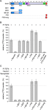
Brd4 PID is an activator of basal and Hexim1-inhibited P-TEFb. (A) Modular domain architecture of murine Brd4. Five different protein constructs were generated that contained the N-terminal bromodomains BD1 and BD2 or the C-terminal P-TEFb interaction domain (PID). Human Brd4 has a total length of 1362 amino acids, whereas mouse Brd4 contains 1400 residues, primarily resulting from an insert of 33 residues at position 1098. Domains are annotated below the diagram with a helical region indicated. (B) Stimulation of P-TEFb activity for RNAPII CTD phosphorylation by Brd4 proteins. The two PID fragments but not the bromodomain containing fragments of Brd4 enhanced the catalytic activity of P-TEFb. PID itself however is not a substrate of P-TEFb. (C) The stimulating effect of PID is even more pronounced upon inhibition of P-TEFb by Hexim1 200-359. Only the PID containing constructs were able to relieve the inhibited state, but not the bromodomains or GST as a control. In contrast, Brd4 PID was unable to activate P-TEFb that has been inhibited with flavopiridol, suggesting a different mechanism of inhibition for the ATP-competitive compound flavopiridol and the cellular regulation factor Hexim1.
Figure 2.
Figure 2.
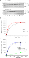
Kinetics of Brd4 PID stimulated P-TEFb activity. (A) SDS-PAGE analysis of P-TEFb-mediated GST-CTD phosphorylation in the absence (upper panel) and presence (lower panel) of PID. The phosphorylation reaction is significantly enhanced in the presence of the PID. (B) Time course experiments revealed a 2-fold higher catalytic efficiency for the P-TEFb-PID complex compared to P-TEFb alone. (C) Inhibition of P-TEFb by Hexim1 is revived by the presence of PID. Even here, the gain in catalytic efficiency is still over basal levels of P-TEFb alone.
Figure 3.
Figure 3.
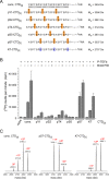
Brd4 PID shares sequence motif similarity to viral Tat proteins. (A) The Brd4 PID contains a surprising similarity in sequence motif assembly to viral Tat proteins, including an RxL motif, a polybasic cluster and a C-terminal leucine motif. (B) Alignment of Brd4 PID sequences from different organisms and generation of four PID mutants. (C) Mutations of the characteristic sequence motifs to alanine impair PID function for P-TEFb stimulation. The RxLAA mutant showed only a small decrease in stimulatory function, whereas mutation of the four arginines (4R4A) fully abrogated the PID stimulatory effect. (D) Upon inhibition by Hexim1 (200-359), the contribution of the PID sequence motifs emerges showing that the polybasic cluster and the leucine motif of PID are necessarily required for P-TEFb activation by Brd4.
Figure 4.
Figure 4.
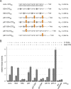
Analysis of Tat sequence motifs for P-TEFb activation. (A) Sequence alignment of Tat proteins from viruses HIV-1, HIV-2, SIV, BIV, JDV and EIAV. A cysteine-rich region, an RxL motif, an arginine-rich motif and a C-terminal leucine motif are conserved in viral Tat proteins. Besides wild type HIV-1 Tat (1-86), three mutants were generated and analyzed for P-TEFb activation. (B) Addition of HIV-1 Tat potently stimulated P-TEFb activity for CTD phosphorylation up to 2.5-fold. Mutation of the KxL motif and the ARM, but to a lesser extent the SL motif, diminished the stimulatory effect of Tat. Yet Tat is not a substrate of P-TEFb as shown by a reaction missing the RNAPII CTD. (C) In the presence of Hexim1, Tat is still able to activate P-TEFb but not as potently as in the absence of the cellular regulator. The KxL motif and the ARM are necessarily required for this activating function, but not the SL motif.
Figure 5.
Figure 5.
Interaction of Brd4 PID with P-TEFb. (A) Isothermal titration calorimetry measurements of Brd4 PID with P-TEFb (Cdk9/CycT1) revealed a dissociation constant of 0.47 μM for this interaction. (B) In contrast, PID showed no binding to CycT1 alone, suggesting Cdk9 as the targeting moiety for Brd4 binding. The thermodynamic parameters of the interaction are listed in Table 1.
Figure 6.
Figure 6.
Brd4 PID increases the activity of P-TEFb for specific CTD substrates. (A) RNAPII CTD substrate peptides used for P-TEFb activity measurements contained three consensus hepta-repeats with either no modifications (cons. CTD[3]), phosphorylation marks continuously set at Tyr1 (pY1-CTD[3]), Ser2 (pS2-CTD[3]), Thr4 (pT4-CTD[3]), Ser5 (pS5-CTD[3]) or Ser7 (pS7-CTD[3]). In addition, a lysine residue was set at position 7 as the most common variation of the consensus CTD (K7-CTD[3]). (B) Substrate preferences of P-TEFb alone or in presence of Brd4 PID. 100 μM of CTD peptides were incubated with 0.2 μM P-TEFb, 1 mM ATP and 2 μM Brd4 PID at 30°C. The recognition of P-TEFb for the various CTD peptides remained unchanged in the P-TEFb-PID complex, but the catalytic activity is significantly enhanced. The phosphorylation efficacy was monitored after 15 min reaction time. (C) ESI-MS analyses of CTD peptides after 4 h incubation with P-TEFb in presence of PID. Up to three phosphorylations were detected for the three repeat containing substrates cons. CTD[3], pS7-CTD[3] and K7-CTD[3].
Figure 7.
Figure 7.
Phosphorylation specificity of P-TEFb in the absence and presence of Brd4 PID. (A) Using the same design of three CTD hepta-repeats as before, continuous alanine mutations were introduced at either Ser2 or Ser5 positions in the consensus CTD (cons. CTD[3]) or in a CTD pre-phosphorylated at position Ser7 (pS7-CTD[3]). In addition, a single Ser7 phosphorylation was set either in the N- or C-terminal repeat of the CTD for its recognition by P-TEFb. (B) Whereas the S2A variants of the CTD both in the non-phosphorylated and the Ser7 pre-phosphorylated form are readily phosphorylated by P-TEFb and the P-TEFb–Brd4 PID complex, the S5A mutants failed being recognized as P-TEFb substrates. The highest stimulatory effect with almost 5-fold increase upon addition of PID is achieved for a substrate peptide containing only one C-terminal Ser7 phosphorylation mark.
Figure 8.
Figure 8.
The P-TEFb–Brd4 PID complex is a Ser5 CTD kinase. (A) Western blot analysis of a full length GST-CTD substrate containing all 52 human repeats revealed that P-TEFb predominantly phosphorylates Ser5 of the CTD. Ser7 is phosphorylated to a lesser extent and only a faint band appeared for Ser2 phosphorylation. Shown is a time course experiment over 8 h using antibodies against pSer2 (3E10, top panel), pSer5 (3E8, middle panel) and pSer7 (4E12, lower panel). (B) In the presence of Brd4 PID, the phosphorylations appear earlier in the time course experiments in line with the stimulatory effect of the PID, but the phosphorylation specificity remains unchanged. Note the migration of the CTD from the hypo-phosphorylated IIa form at the beginning of the reaction (5 min) to the hyper-phosphorylated IIo form with the highest levels after 120 min. (C) Input control of the GST-CTD[52] substrate. A coomassie stained SDS-PAGE analysis of the full length hepta-repeat region of human RNAPII CTD is shown, indicating minor fractions of leaky GST but no major hepta-repeat truncations.

References

    1. Adelman K., Lis J.T. Promoter-proximal pausing of RNA polymerase II: emerging roles in metazoans. Nat. Rev. Genet. 2012;13:720–731.. - PMC - PubMed
    1. Zhou Q., Li T., Price D.H. RNA polymerase II elongation control. Annu. Rev. Biochem. 2012;81:119–143. - PMC - PubMed
    1. Eick D., Geyer M. The RNA polymerase II carboxy-terminal domain (CTD) code. Chem. Rev. 2013;113:8456–8490. - PubMed
    1. Prasanth K.V., Camiolo M., Chan G., Tripathi V., Denis L., Nakamura T., Hübner M.R., Spector D.L. Nuclear organization and dynamics of 7SK RNA in regulating gene expression. Mol. Biol. Cell. 2010;21:4184–4196. - PMC - PubMed
    1. Lebars I., Martinez-Zapien D., Durand A., Coutant J., Kieffer B., Dock-Bregeon A.C. HEXIM1 targets a repeated GAUC motif in the riboregulator of transcription 7SK and promotes base pair rearrangements. Nucleic Acids Res. 2010;38:7749–7763. - PMC - PubMed

Publication types

MeSH terms