

Disease-associated CAG·CTG triplet repeats expand rapidly in non-dividing mouse cells, but cell cycle arrest is insufficient to drive expansion
- PMID: 24860168
- PMCID: PMC4066746
- DOI: 10.1093/nar/gku285
Disease-associated CAG·CTG triplet repeats expand rapidly in non-dividing mouse cells, but cell cycle arrest is insufficient to drive expansion
Abstract
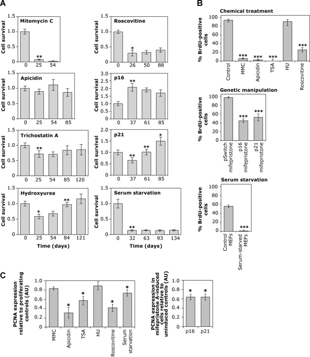
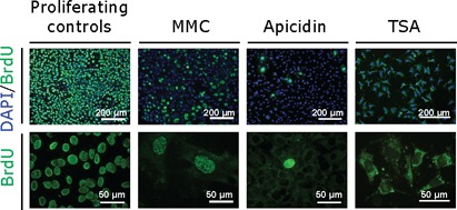
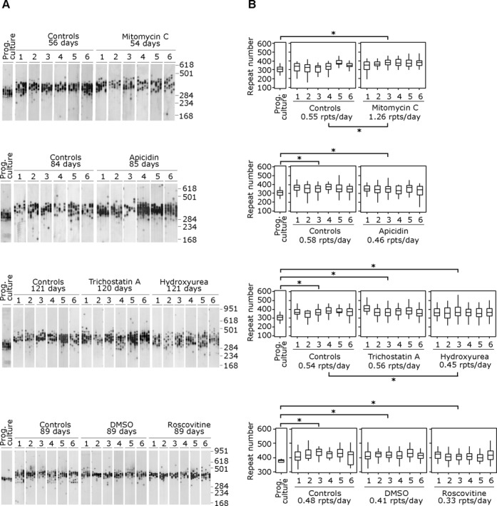
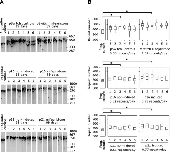
Genetically unstable expanded CAG·CTG trinucleotide repeats are causal in a number of human disorders, including Huntington disease and myotonic dystrophy type 1. It is still widely assumed that DNA polymerase slippage during replication plays an important role in the accumulation of expansions. Nevertheless, somatic mosaicism correlates poorly with the proliferative capacity of the tissue and rates of cell turnover, suggesting that expansions can occur in the absence of replication. We monitored CAG·CTG repeat instability in transgenic mouse cells arrested by chemical or genetic manipulation of the cell cycle and generated unequivocal evidence for the continuous accumulation of repeat expansions in non-dividing cells. Importantly, the rates of expansion in non-dividing cells were at least as high as those of proliferating cells. These data are consistent with a major role for cell division-independent expansion in generating somatic mosaicism in vivo. Although expansions can accrue in non-dividing cells, we also show that cell cycle arrest is not sufficient to drive instability, implicating other factors as the key regulators of tissue-specific instability. Our data reveal that de novo expansion events are not limited to S-phase and further support a cell division-independent mutational pathway.
© The Author(s) 2014. Published by Oxford University Press on behalf of Nucleic Acids Research.
Figures
Similar articles
-
Age-, tissue- and length-dependent bidirectional somatic CAG•CTG repeat instability in an allelic series of R6/2 Huntington disease mice.Neurobiol Dis. 2015 Apr;76:98-111. doi: 10.1016/j.nbd.2015.01.004. Epub 2015 Feb 3. Neurobiol Dis. 2015. PMID: 25662336
-
Mouse tissue culture models of unstable triplet repeats: in vitro selection for larger alleles, mutational expansion bias and tissue specificity, but no association with cell division rates.Hum Mol Genet. 2001 Apr 1;10(8):845-54. doi: 10.1093/hmg/10.8.845. Hum Mol Genet. 2001. PMID: 11285250
-
Pms2 is a genetic enhancer of trinucleotide CAG.CTG repeat somatic mosaicism: implications for the mechanism of triplet repeat expansion.Hum Mol Genet. 2004 Aug 15;13(16):1815-25. doi: 10.1093/hmg/ddh186. Epub 2004 Jun 15. Hum Mol Genet. 2004. PMID: 15198993
-
The unstable trinucleotide repeat story of major psychosis.Am J Med Genet. 2000 Spring;97(1):77-97. doi: 10.1002/(sici)1096-8628(200021)97:1<77::aid-ajmg11>3.0.co;2-3. Am J Med Genet. 2000. PMID: 10813808 Review.
-
DNA trinucleotide repeat expansion in neuropsychiatric patients.Med Sci Monit. 2003 Sep;9(9):RA237-45. Med Sci Monit. 2003. PMID: 12960939 Review.
Cited by
-
Huntington disease: new insights into molecular pathogenesis and therapeutic opportunities.Nat Rev Neurol. 2020 Oct;16(10):529-546. doi: 10.1038/s41582-020-0389-4. Epub 2020 Aug 14. Nat Rev Neurol. 2020. PMID: 32796930 Review.
-
The Repeat Expansion Diseases: The dark side of DNA repair.DNA Repair (Amst). 2015 Aug;32:96-105. doi: 10.1016/j.dnarep.2015.04.019. Epub 2015 Apr 30. DNA Repair (Amst). 2015. PMID: 26002199 Free PMC article. Review.
-
Variant repeats within the DMPK CTG expansion protect function in myotonic dystrophy type 1.Neurol Genet. 2020 Aug 12;6(5):e504. doi: 10.1212/NXG.0000000000000504. eCollection 2020 Oct. Neurol Genet. 2020. PMID: 32851192 Free PMC article.
-
Sex contribution to average age at onset of Huntington's disease depends on the number of (CAG)n repeats.Sci Rep. 2024 Jul 8;14(1):15729. doi: 10.1038/s41598-024-64105-5. Sci Rep. 2024. PMID: 38977715 Free PMC article.
-
Break-induced replication links microsatellite expansion to complex genome rearrangements.Bioessays. 2017 Aug;39(8):10.1002/bies.201700025. doi: 10.1002/bies.201700025. Epub 2017 Jun 16. Bioessays. 2017. PMID: 28621832 Free PMC article. Review.
References
-
- Gomes-Pereira M., Monckton D.G. Chemical modifiers of unstable expanded simple sequence repeats: what goes up, could come down. Mutat. Res. 2006;598:15–34. - PubMed
-
- Monckton D.G., Wong L.J., Ashizawa T., Caskey C.T. Somatic mosaicism, germline expansions, germline reversions and intergenerational reductions in myotonic dystrophy males: small pool PCR analyses. Hum. Mol. Genet. 1995;4:1–8. - PubMed
-
- Higham C.F., Morales F., Cobbold C.A., Haydon D.T., Monckton D.G. High levels of somatic DNA diversity at the myotonic dystrophy type 1 locus are driven by ultra-frequent expansion and contraction mutations. Hum. Mol. Genet. 2012;21:2450–2463. - PubMed
-
- Ashizawa T., Dube J.R., Harat Y. Somatic instability of CTG repeat in myotonic dystrophy. Neurology. 1993;43:2674–2678. - PubMed
Publication types
MeSH terms
Grants and funding
LinkOut - more resources
Full Text Sources
Other Literature Sources
