. 2014 Aug 15;592(16):3443-61.
doi: 10.1113/jphysiol.2014.275065. Epub 2014 May 23.

Native store-operated calcium channels are functionally expressed in mouse spinal cord dorsal horn neurons and regulate resting calcium homeostasis

Affiliations

Native store-operated calcium channels are functionally expressed in mouse spinal cord dorsal horn neurons and regulate resting calcium homeostasis

Jingsheng Xia et al. J Physiol. .

Abstract

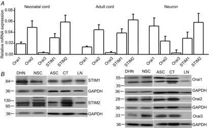
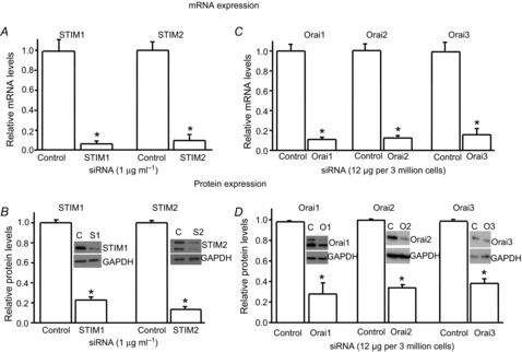
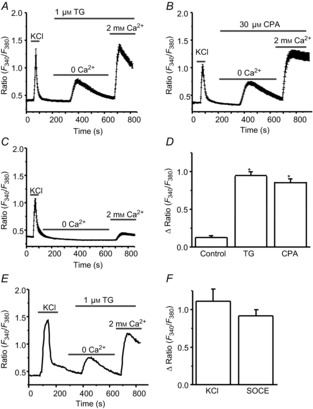
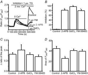
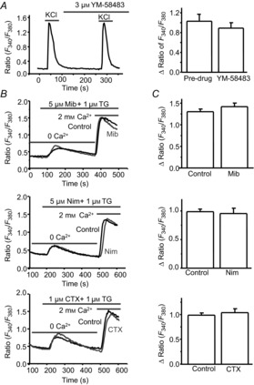
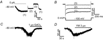
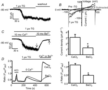
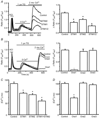
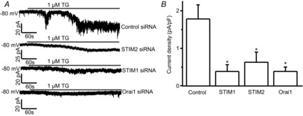
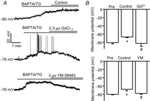
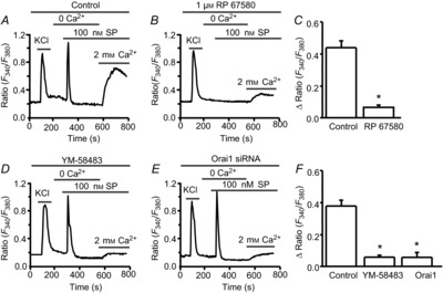
Store-operated calcium channels (SOCs) are calcium-selective cation channels that mediate calcium entry in many different cell types. Store-operated calcium entry (SOCE) is involved in various cellular functions. Increasing evidence suggests that impairment of SOCE is responsible for numerous disorders. A previous study demonstrated that YM-58483, a potent SOC inhibitor, strongly attenuates chronic pain by systemic or intrathecal injection and completely blocks the second phase of formalin-induced spontaneous nocifensive behaviour, suggesting a potential role of SOCs in central sensitization. However, the expression of SOCs, their molecular identity and function in spinal cord dorsal horn neurons remain elusive. Here, we demonstrate that SOCs are expressed in dorsal horn neurons. Depletion of calcium stores from the endoplasmic reticulum (ER) induced large sustained calcium entry, which was blocked by SOC inhibitors, but not by voltage-gated calcium channel blockers. Depletion of ER calcium stores activated inward calcium-selective currents, which was reduced by replacing Ca(2+) with Ba(2+) and reversed by SOC inhibitors. Using the small inhibitory RNA knockdown approach, we identified both STIM1 and STIM2 as important mediators of SOCE and SOC current, and Orai1 as a key component of the Ca(2+) release-activated Ca(2+) channels in dorsal horn neurons. Knockdown of STIM1, STIM2 or Orai1 decreased resting Ca(2+) levels. We also found that activation of neurokinin 1 receptors led to SOCE and activation of SOCs produced an excitatory action in dorsal horn neurons. Our findings reveal that a novel SOC signal is present in dorsal horn neurons and may play an important role in pain transmission.


Figures

Figure 1
Figure 1. Store-operated calcium channels (SOCs) are expressed in spinal cord dorsal horn neurons
A, mRNA levels of STIM1, STIM2, Orai1, Orai2 and Orai3 in neonatal and adult spinal cord tissue and acutely dissociated dorsal horn neurons by quantitative PCR (normalized to GAPDH). Values represent mean ± SEM, = 4 samples each. B, protein levels of STIM1, STIM2, Orai1, Orai2 and Orai3 in dorsal horn neurons (DHN), neonatal spinal cord (NSC), adult spinal cord (ASC), the cortex (CT) and lymph nodes (LN) by Western blot analysis.
Figure 2
Figure 2. Expression of the SOC family is significantly decreased by transfection of target-specific siRNAs in dorsal horn neurons
A and B, effects of specific siRNAs against STIM1 or STIM2 on mRNA levels (A) and protein levels (B) of STIM1 and STIM2, respectively (= 4–5 samples). C and D, effects of specific siRNAs against Orai1, Orai2 or Orai3 on mRNA levels (C) and protein levels (D) of Orai1, Orai2 and Orai3, respectively (= 4 samples each). The mRNA and protein levels were normalized to control (non-target siRNA). Values represent mean ± SEM; *P < 0.05, compared with control by Student's t test.
Figure 3
Figure 3. Depletion of endoplasmic reticulum Ca2+ stores by thapsigargin (TG) and cyclopiazonic acid (CPA) induces calcium responses in cultured dorsal horn neurons
A, TG-induced calcium entry (= 53 neurons). B, CPA-induced calcium entry (= 57 neurons). C, addition of 2 mm Ca2+-induced calcium entry in the absence of TG or CPA (= 39 neurons). D, summary of TG- and CPA-induced calcium influx. E, KCl- and TG-induced calcium entry (= 19). F, summary of KCl- and TG-induced calcium influx. Values represent mean ± SEM; *P < 0.05, compared with control by one-way ANOVA.
Figure 4
Figure 4. TG-induced calcium entry is inhibited by SOC inhibitors
A, representative TG-induced calcium responses recorded in neurons pretreated with 2-APB, YM-58483 or GdCl3; B, summary of the effects of 2-APB (= 32 neurons), YM-58483 (=  41 neurons) and GdCl3 (= 45 neurons) on TG-induced calcium entry. C and D, summary of the effects of 2-APB, YM-58483 and GdCl3 on TG-induced calcium release presented as the peak amplitude (C) or as area (D). Values represent mean ± SEM; *P < 0.05, compared with control by one-way ANOVA.
Figure 5
Figure 5. CPA-induced calcium entry is attenuated by SOC inhibitors
A, effect of GdCl3 on CPA-induced calcium influx. Left, representative examples; right, summary of the effect of GdCl3 (= 15 neurons). B, effect of YM-58483 on CPA-induced calcium influx. Left, representative examples; right, summary of the effect of YM-58483 (= 18 neurons). C, effect of 2-APB on CPA-induced calcium influx. Left, representative examples; right, summary of the effect of 2-APB (= 11 neurons). Values represent mean ± SEM; *P < 0.05, compared with control by one-way ANOVA.
Figure 6
Figure 6. Store-operated calcium entry (SOCE) is independent of voltage-gated calcium channels (VGCCs)
A, effect of YM-58483 on KCl-induced calcium response (= 15 neurons). Left, representative 60 mm KCl-induced calcium responses; right, summary of the effect of YM-58483. B, representative TG-induced calcium responses recorded in neurons pretreated with mibefradil (Mib), nimodipine (Nim) or ω-conotoxin MVIIC (CTX). C, summary of the effects of mibefradil (= 50 neurons), nimodipine (= 45 neurons) and ω-conotoxin MVIIC (= 32 neurons) on TG-induced SOCE. Values represent mean ± SEM; *P < 0.05, compared with control by Student's t test.
Figure 7
Figure 7. Depletion of Ca2+ stores by BAPTA induces SOC currents in dorsal horn neurons
A and B, effects of replacing 10 mm Na+ with 10 mm Cs+ or removing Ca2+ on BAPTA-induced currents recorded with a gap-free protocol (A) and a step protocol (B). (1) normal Tyrode's solution; (2) replacing 10 mm NaCl with CsCl; (3) removing Ca2+ from (2). C and D, BAPTA-induced currents were attenuated by GdCl3 (C) or YM-58483 (D).
Figure 8
Figure 8. BAPTA/TG-induced SOC currents are mediated by CRAC channels in dorsal horn neurons
A and B, BAPTA/TG-induced calcium currents recorded with the gap-free protocol (A) and with a ramp protocol (B). C, effect of replacing 10 mm Ca2+ with 10 mm Ba2+ on BAPTA/TG-induced current recorded with the gap-free protocol. Left, a representative BAPTA/TG-induced current recorded in 10 mm Ca2+ or 10 mm Ba2+; right, summary of BAPTA/TG-induced Ca2+ current and Ba2+ current (= 9 neurons). D, effect of replacing 2 mm Ca2+ with 2 mm Ba2+ on TG-induced SOCE. Left, representative TG-induced Ca2+ or Ba2+ entry; right, summary of TG-induced Ca2+ entry (= 52 neurons) and Ba2+ entry (= 40 neurons). Arrows indicate where current amplitudes were measured. Values represent mean ± SEM; *P < 0.05, compared by Student's t test.
Figure 9
Figure 9. SOCs are functional in lamina I/II neurons from spinal cord slices of adult mice
A–C, representative BAPTA/TG-induced SOC currents recorded in neurons from slices and from cultures, which are reduced by 2-APB (A), GdCl3 (B) or YM-58483 (C). D–F, summary of inhibition of BAPTA/TG-induced SOC currents by 2-APB (= 7–9 neurons), GdCl3 (= 8–11 neurons) or YM-58483 (= 9–12 neurons). Arrows indicate where current amplitudes were measured. Values represent mean ± SEM; *P < 0.05, compared with control (pre-drug, Pre) by paired Student's t test.
Figure 10
Figure 10. Orai1 is required, and STIM1 and STIM2 are important for SOCE and resting calcium homeostasis in dorsal horn neurons
A, effects of specific siRNAs against STIMs on TG-induced SOCE. Left, representative examples of TG-induced calcium responses recorded in neurons transfected with control siRNA or targeting siRNAs against STIMs; right, summary of effects of control siRNA (= 78 neurons), STIM1 siRNA (= 77 neurons), STIM2 siRNA (= 69 neurons) and mixed siRNAs against STIM1 and STIM2 (= 32 neurons), respectively. B, effects of specific siRNAs against Orai members on TG-induced SOCE. Left, representative examples of TG-induced calcium responses recorded in neurons transfected with control siRNA or targeting siRNAs against Orai members; right, summary of the effects of control siRNA (= 112 neurons), Orai1 siRNA (= 76 neurons), Orai2 siRNA (= 40 neurons) or Orai3 siRNA (= 42 neurons). C, effects of specific siRNAs against STIMs on the resting calcium concentration measured in neurons transfected with control siRNA (= 99 neurons), STIM1 siRNA (= 99 neurons), STIM2 siRNA (= 67 neurons) or mixed siRNAs against STIM1 and STIM2 (= 45). D, effects of specific siRNAs against Orai members on the resting calcium concentration measured in neurons transfected with control siRNA (= 329 neurons), Orai1 siRNA (= 109 neurons), Orai2 siRNA (= 114 neurons) or Orai3 siRNA (= 98 neurons). Values represent mean ± SEM, *P < 0.05, compared with control by one-way ANOVA.
Figure 11
Figure 11. SOC currents are mediated by STIM1, STIM2 and Orai1
Effects of specific siRNAs against STIM1, STIM2 or Orai1 on BAPTA/TG-induced calcium currents. A, representative BAPTA/TG-induced calcium currents recorded with the gap-free protocol in neurons transfected with control siRNA, targeting siRNAs. B, summary of the effects of control siRNA (= 25 neurons), STIM1 siRNAs (= 16 neurons), STIM2 siRNA (= 18 neurons) and Orai1 siRNA (= 18 neurons), respectively. Values represent mean ± SEM; *P < 0.05, compared with control by one-way ANOVA.
Figure 12
Figure 12. Activation of SOCs induces membrane depolarization in dorsal horn neurons
A, representative examples of BAPTA/TG-induced depolarization treated with control, 0.3 μm GdCl3 or 3 μm YM-58483. B, summary of the effects of GdCl3 or YM-58483 on BAPTA/TG-induced changes in membrane potential. Values represent mean ± SEM; = 5–9 neurons each; *P < 0.05 compared with pre (before TG application), #P < 0.05 compared with control by one-way ANOVA.
Figure 13
Figure 13. Activation of NK1 receptors results in SOCE in dorsal horn neurons
A and B, representative examples of substance P (SP)-induced Ca2+ responses in the absence (A), or in the presence of 1 μm RP 67580 (B). C, summary of the effect of RP 67580 on SP-induced SOCE. D and E, representative examples of SP-induced Ca2+ responses in the presence of YM-58483 (D), or after knockdown of Orai1 (E). F, summary of the effects of 3 μm YM-58483 and knockdown of Orai1 on SP-induced SOCE. Values represent mean ± SEM; = 42–54 neurons each; *P < 0.05, compared with control by Student's t test (C) or one-way ANOVA (F).

Similar articles

Cited by

References

    1. Baba A, Yasui T, Fujisawa S, Yamada RX, Yamada MK, Nishiyama N, Matsuki N. Ikegaya Y. Activity-evoked capacitative Ca2+ entry: implications in synaptic plasticity. J Neurosci. 2003;23:7737–7741. - PMC - PubMed
    1. Bakowski D. Parekh AB. Voltage-dependent Ba2+ permeation through store-operated CRAC channels: implications for channel selectivity. Cell Calcium. 2007;42:333–339. - PubMed
    1. Berna-Erro A, Braun A, Kraft R, Kleinschnitz C, Schuhmann MK, Stegner D, Wultsch T, Eilers J, Meuth SG, Stoll G. Nieswandt B. STIM2 regulates capacitive Ca2+ entry in neurons and plays a key role in hypoxic neuronal cell death. Sci Signal. 2009;2:ra67. - PubMed
    1. Bezprozvanny I. Hayden MR. Deranged neuronal calcium signaling and Huntington disease. Biochem Biophys Res Commun. 2004;322:1310–1317. - PubMed
    1. Bird GS, DeHaven WI, Smyth JT. Putney JW., Jr Methods for studying store-operated calcium entry. Methods. 2008;46:204–212. - PMC - PubMed

Publication types

LinkOut - more resources