. 2014:2014:954502.
doi: 10.1155/2014/954502. Epub 2014 Apr 17.

Protective effect of astaxanthin on liver fibrosis through modulation of TGF-β1 expression and autophagy

Affiliations

Protective effect of astaxanthin on liver fibrosis through modulation of TGF-β1 expression and autophagy

Miao Shen et al. Mediators Inflamm. 2014.

Abstract

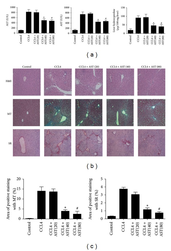
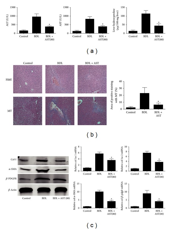
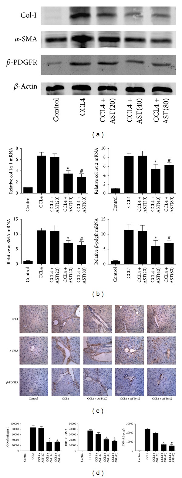
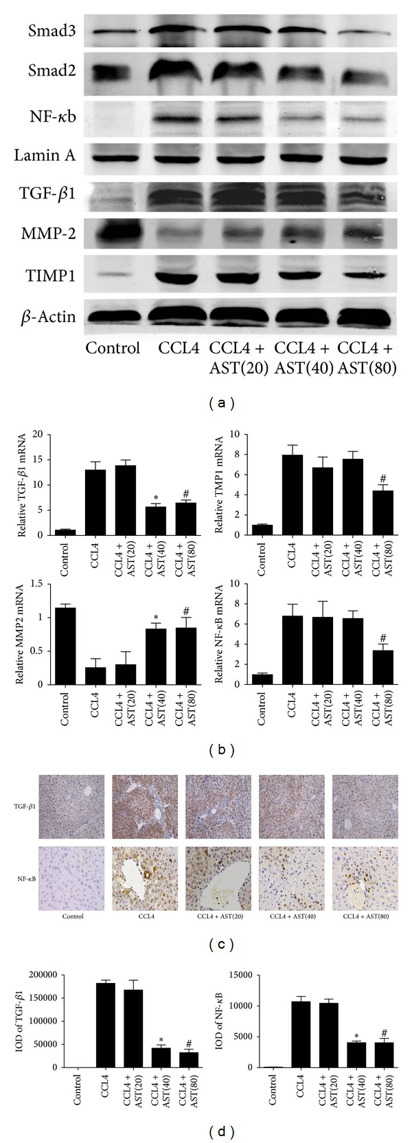
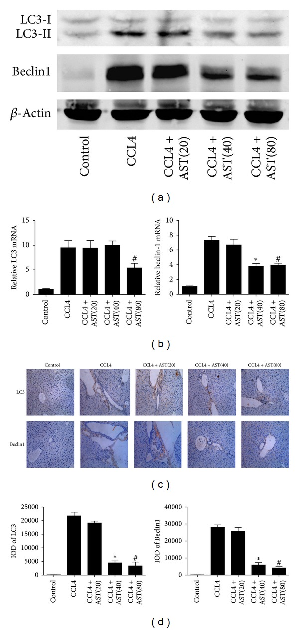
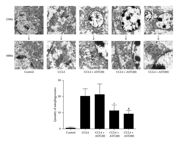
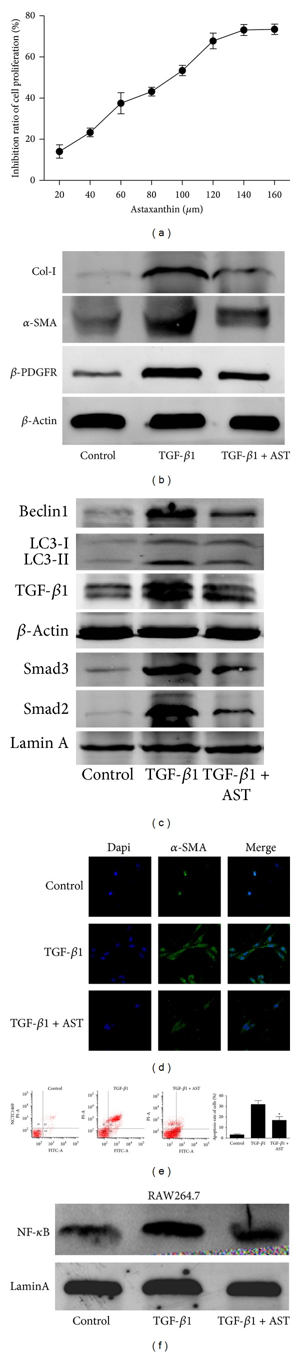
Liver fibrosis is a common pathway leading to cirrhosis and a worldwide clinical issue. Astaxanthin is a red carotenoid pigment with antioxidant, anticancer, and anti-inflammatory properties. The aim of this study was to investigate the effect of astaxanthin on liver fibrosis and its potential protective mechanisms. Liver fibrosis was induced in a mouse model using CCL4 (intraperitoneal injection, three times a week for 8 weeks), and astaxanthin was administered everyday at three doses (20, 40, and 80 mg/kg). Pathological results indicated that astaxanthin significantly improved the pathological lesions of liver fibrosis. The levels of alanine aminotransferase aspartate aminotransferase and hydroxyproline were also significantly decreased by astaxanthin. The same results were confirmed in bile duct liagtion, (BDL) model. In addition, astaxanthin inhibited hepatic stellate cells (HSCs) activation and formation of extracellular matrix (ECM) by decreasing the expression of NF-κB and TGF-β1 and maintaining the balance between MMP2 and TIMP1. In addition, astaxanthin reduced energy production in HSCs by downregulating the level of autophagy. These results were simultaneously confirmed in vivo and in vitro. In conclusion, our study showed that 80 mg/kg astaxanthin had a significant protective effect on liver fibrosis by suppressing multiple profibrogenic factors.


Figures

Figure 1
Figure 1
Effect of astaxanthin on CCL4-induced liver fibrosis. (a) Astaxanthin decreased the level of ALT, AST, and hydroxyproline with the doses of 40 mg/kg and 80 mg/kg. Data are expressed as mean ± SD (n = 7, *P < 0.05 for CCL4 + AST(40) versus CCL4, # P < 0.05 for CCL4 + AST(80) versus CCL4). (b) Astaxanthin (40 mg/kg and 80 mg/kg) ameliorated pathological change showed by H&E, Masson's trichrome (MT), and sirius red staining (original magnification: ×200). (c) The areas of positive staining with MT and SR were analyzed by Image-pro Plus 6.0. There existed a significant decrease with astaxanthin (40 mg/kg and 80 mg/kg) treatment (n = 7, *P < 0.05 for CCL4 + AST(40) versus CCL4, # P < 0.05 for CCL4 + AST(80) versus CCL4).
Figure 2
Figure 2
Effect of astaxanthin on BDL-induced liver fibrosis. (a) Astaxanthin decreased the level of ALT, AST, and hydroxyproline with the dose of 80 mg/kg. Data are expressed as mean ± SD (n = 10, *P < 0.05 for BDL + AST(80) versus BDL). (b) Astaxanthin (80 mg/kg) ameliorated pathological change showed by H&E and Masson's trichrome (MT) (original magnification: ×200). The areas of positive staining with MT were analyzed by Image-pro Plus 6.0. There existed a significant decrease with astaxanthin (80 mg/kg) treatment (n = 10, *P < 0.05 for BDL + AST(80) versus BDL). (c) The analysis of western blotting and real-time PCR showed that astaxanthin obviously decreased the expression of α-SMA, β-pdfgr, and collagen I with the doses of 80 mg/kg in BDL model (n = 3, *P < 0.05 for BDL + AST(80) versus BDL).
Figure 3
Figure 3
Effect of astaxanthin on the activation of HSCs. (a) The analysis of western blotting showed that astaxanthin obviously decreased the expression of α-SMA, β-pdfgr, and collagen I with the doses of 40 mg/kg and 80 mg/kg. (b) The mRNA levels of collagen I  α1, collagen I  α2, α-SMA, and β-pdgfr were significantly downregulated by astaxanthin (40 mg/kg and 80 mg/kg). Data are expressed as mean ± SD (n = 7, *P < 0.05 for CCL4 + AST(40) versus CCL4, # P < 0.05 for CCL4 + AST(80) versus CCL4). (c) The areas of positive cells of α-SMA, β-pdfgr, and collagen I were diminished by astaxanthin (40 mg/kg and 80 mg/kg), showed by immunohistochemistry staining (original magnification: ×200). (d) The IODs of α-SMA, β-pdfgr, and collagen I were analyzed by Image-pro Plus 6.0. There existed a significant decrease with astaxanthin (40 mg/kg and 80 mg/kg) treatment (n = 7, *P < 0.05 for CCL4 + AST(40) versus CCL4, # P < 0.05 for CCL4 + AST(80) versus CCL4).
Figure 4
Figure 4
Effects of astaxanthin on expression of TGF-β1, MMP2, TIMP1, and NF-κb. (a) The analysis of western blotting showed that astaxanthin decreased the expression of TGF-β1, TIMP1, and NF-κb and, contrarily, increased the expression of MMP2 compared to CCL4 group. (b) The mRNA level of TGF-β1 was decreased by astaxanthin (40 mg/kg and 80 mg/kg), and the mRNA levels of TIMP1 and NF-κb were decreased with the dose of 80 mg/kg. However, the mRNA levels of MMP2 were increased by astaxanthin (40 mg/kg and 80 mg/kg) compared to CCL4 group. Data are expressed as mean ± SD (n = 7, *P < 0.05 for CCL4 + AST(40) versus CCL4, # P < 0.05 for CCL4 + AST(80) versus CCL4). (c) The area of positive cells of TGF-β1 was significantly decreased by astaxanthin (40 mg/kg and 80 mg/kg) stained by immunohistochemistry (original magnification: ×200). The expression of NF-κB in nuclei decreased obviously with astaxanthin (40 mg/kg and 80 mg/kg) treatment (original magnification: ×400). (d) The IODs of TGF-β1 and NF-κb were analyzed by Image-pro Plus 6.0. There existed a significant decrease with astaxanthin (40 mg/kg and 80 mg/kg) treatment (n = 7, *P < 0.05 for CCL4 + AST(40) versus CCL4, # P < 0.05 for CCL4 + AST(80) versus CCL4).
Figure 5
Figure 5
Effect of astaxanthin on the regulation of autophagy in HSCs. (a) The analysis of western blotting showed that astaxanthin (40 mg/kg and 80 mg/kg) obviously decreased the expression of LC3-II and beclin-1. (b) The mRNA levels of beclin-1 were decreased by astaxanthin (40 mg/kg and 80 mg/kg) compared to CCL4 group, and the level of LC3 was decreased only with the dose of 80 mg/kg. Data are expressed as mean ± SD (n = 7, *P < 0.05 for CCL4 + AST(40) versus CCL4, # P < 0.05 for CCL4 + AST(80) versus CCL4). (c) The areas of positive cells of LC3 and beclin-1, mainly expressed in HSCs as showed, were visibly diminished by astaxanthin (40 mg/kg and 80 mg/kg) (original magnification: ×200). (d) The IODs of LC3 and beclin-1 were analyzed by Image-pro Plus 6.0. There existed a significant decrease with astaxanthin (40 mg/kg and 80 mg/kg) treatment (n = 7, *P < 0.05 for CCL4 + AST(40) versus CCL4, # P < 0.05 for CCL4 + AST(80) versus CCL4).
Figure 6
Figure 6
Effect of astaxanthin on the formation of autophagosome. As shown in picture, the amount of autophagosome obviously increased compared to control group. After astaxanthin (40 mg/kg and 80 mg/kg) treated, the amount of autophagosome significantly decreased, and the ultrastructure of cells exhibited more integrated (autophagosome was indicated with “→”) (original magnification: ×2500 and ×5000).
Figure 7
Figure 7
Effect of astaxanthin on HSC-T6, NCTC1469, and RAW264.7 cells. (a) The proliferation of HSC-T6 was detected by MTT. (b) The levels of α-SMA, β-pdfgr, and collagen I were detected by western blot. (c) The levels of beclin-1, LC3, TGF-β1, Smad2, and Smad3 were detected by western blot. (d) The expression of α-SMA in HSC-T6 cells was detected by immunofluorescence. (e) The apoptosis of NCTC1469 cells was detected by flow cytometry (n = 3, *P < 0.05 for TGF-β1 + AST(40) versus TGF-β1). (f) The result of western blot showed that astaxanthin decreased the expression of NF-κB in RAW264.7 cells.

References

    1. Wynn TA. Cellular and molecular mechanisms of fibrosis. Journal of Pathology. 2008;214(2):199–210. - PMC - PubMed
    1. Kisseleva T, Brenner DA. Role of hepatic stellate cells in fibrogenesis and the reversal of fibrosis. Journal of Gastroenterology and Hepatology. 2007;22(1):S73–S78. - PubMed
    1. Jiang JX, Torok NJ. Liver injury and the activation of the hepatic myofibroblasts. Current Pathobiology Reports. 2013;1(3):215–223. - PMC - PubMed
    1. Tacke F, Weiskirchen R. Update on hepatic stellate cells: pathogenic role in liver fibrosis and novel isolation techniques. Expert Review of Gastroenterology and Hepatology. 2012;6(1):67–80. - PubMed
    1. Baghy K, Iozzo RV, Kovalszky I. Decorin-TGFbeta axis in hepatic fibrosis and cirrhosis. Journal of Histochemistry & Cytochemistry. 2012;60(4):262–268. - PMC - PubMed

Publication types