

Regulation of neurotrophin receptor (Trk) signaling: suppressor of cytokine signaling 2 (SOCS2) is a new player
- PMID: 24860421
- PMCID: PMC4030161
- DOI: 10.3389/fnmol.2014.00039
Regulation of neurotrophin receptor (Trk) signaling: suppressor of cytokine signaling 2 (SOCS2) is a new player
Abstract
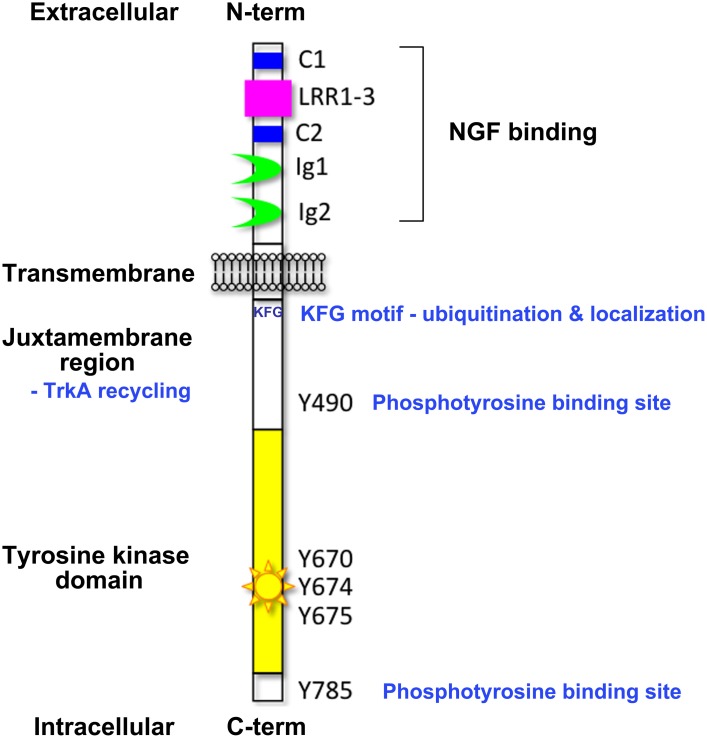
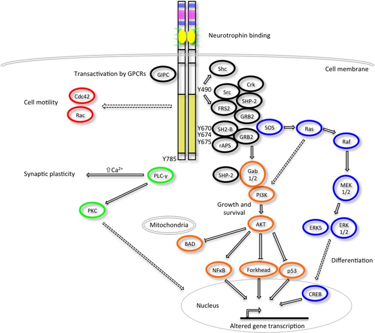
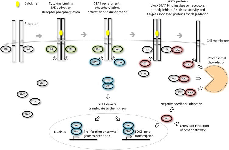
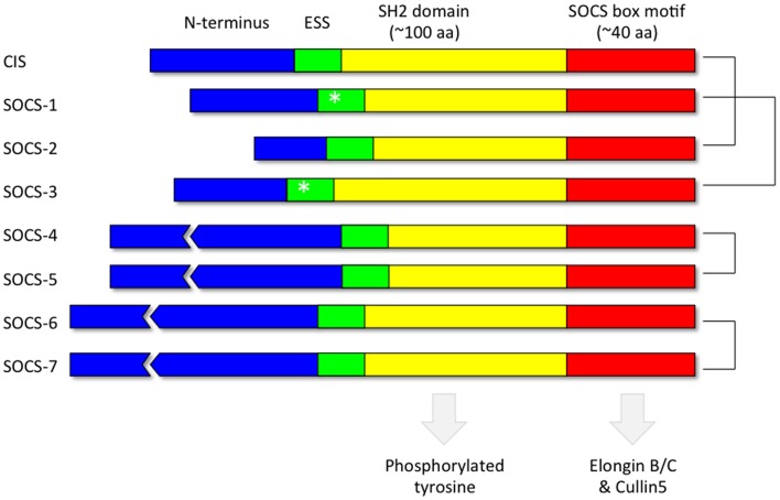
The classic neurotrophins Nerve Growth Factor (NGF), Brain Derived Neurotrophic Factor (BDNF) and Neurotrophins NT-3 and NT-4 are well known to regulate various aspects of neuronal differentiation, survival and growth. They do this by binding to their cognate receptors, members of the Tropomyosin-related kinase (Trk) receptor tyrosine kinase family, namely TrkA, TrkB, and TrkC. These receptors are then internalized and localized to different cellular compartments, where signal transduction occurs. Conversely, members of the suppressor of cytokine signaling (SOCS) family are best known as negative regulators of signaling via the JAK/STAT pathway. Some members of the family, and in particular SOCS2, have roles in the nervous system that at least partially overlap with that of neurotrophins, namely neuronal differentiation and neurite outgrowth. Recent evidence suggests that SOCS2 is a novel regulator of NGF signaling, altering TrkA cellular localization and downstream signaling to affect neurite growth but not neuronal survival. This review first discusses regulation of Trk receptor signaling, followed by the role of SOCS2 in the nervous system and finishes with a discussion of possible mechanisms by which SOCS2 may regulate TrkA function.
Keywords: DRG neurons; NGF; PC12 cells; SOCS-2; Trk receptors; neurite outgrowth; signal transduction; ubiquitin.
Figures
Similar articles
-
Differential dependency of cutaneous mechanoreceptors on neurotrophins, trk receptors, and P75 LNGFR.Dev Biol. 1997 Oct 1;190(1):94-116. doi: 10.1006/dbio.1997.8658. Dev Biol. 1997. PMID: 9331334
-
Binding of neurotrophin-3 to p75LNGFR, TrkA, and TrkB mediated by a single functional epitope distinct from that recognized by trkC.J Biol Chem. 1996 Mar 8;271(10):5623-7. doi: 10.1074/jbc.271.10.5623. J Biol Chem. 1996. PMID: 8621424
-
A novel role of suppressor of cytokine signaling-2 in the regulation of TrkA neurotrophin receptor biology.J Neurochem. 2014 May;129(4):614-27. doi: 10.1111/jnc.12671. Epub 2014 Mar 4. J Neurochem. 2014. PMID: 24484474
-
Neurotrophins in cultured cells from periodontal tissues.J Periodontol. 2003 Jan;74(1):76-84. doi: 10.1902/jop.2003.74.1.76. J Periodontol. 2003. PMID: 12593600 Review.
-
The Trk family of neurotrophin receptors.J Neurobiol. 1994 Nov;25(11):1386-403. doi: 10.1002/neu.480251107. J Neurobiol. 1994. PMID: 7852993 Review.
Cited by
-
Neurotrophin Signaling and Stem Cells-Implications for Neurodegenerative Diseases and Stem Cell Therapy.Mol Neurobiol. 2017 Nov;54(9):7401-7459. doi: 10.1007/s12035-016-0214-7. Epub 2016 Nov 5. Mol Neurobiol. 2017. PMID: 27815842 Review.
-
The role of rab proteins in neuronal cells and in the trafficking of neurotrophin receptors.Membranes (Basel). 2014 Oct 6;4(4):642-77. doi: 10.3390/membranes4040642. Membranes (Basel). 2014. PMID: 25295627 Free PMC article. Review.
-
Role of SOCS and VHL Proteins in Neuronal Differentiation and Development.Int J Mol Sci. 2023 Feb 15;24(4):3880. doi: 10.3390/ijms24043880. Int J Mol Sci. 2023. PMID: 36835292 Free PMC article. Review.
-
Shexiang Baoxin Pill, a Formulated Chinese Herbal Mixture, Induces Neuronal Differentiation of PC12 Cells: A Signaling Triggered by Activation of Protein Kinase A.Front Pharmacol. 2019 Oct 9;10:1130. doi: 10.3389/fphar.2019.01130. eCollection 2019. Front Pharmacol. 2019. PMID: 31649530 Free PMC article.
-
Stingless Bee Honey Improves Spatial Memory in Mice, Probably Associated with Brain-Derived Neurotrophic Factor (BDNF) and Inositol 1,4,5-Triphosphate Receptor Type 1 (Itpr1) Genes.Evid Based Complement Alternat Med. 2019 Dec 2;2019:8258307. doi: 10.1155/2019/8258307. eCollection 2019. Evid Based Complement Alternat Med. 2019. PMID: 31885664 Free PMC article.
References
Publication types
LinkOut - more resources
Full Text Sources
Other Literature Sources
Research Materials