. 2014 May 5:5:389.
doi: 10.3389/fpsyg.2014.00389. eCollection 2014.

Multisensory and modality specific processing of visual speech in different regions of the premotor cortex

Affiliations

Multisensory and modality specific processing of visual speech in different regions of the premotor cortex

Daniel E Callan et al. Front Psychol. .

Abstract

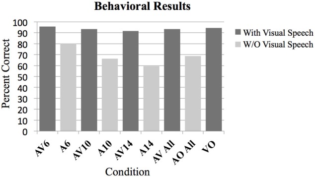
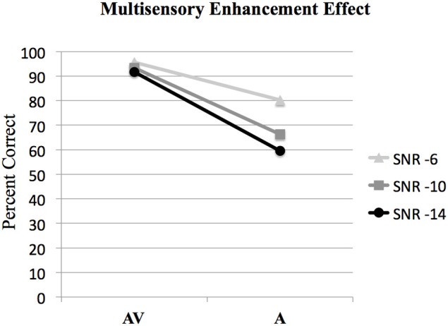
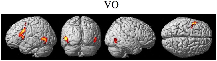
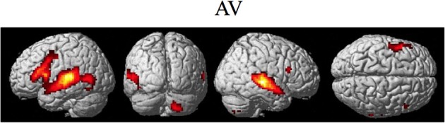
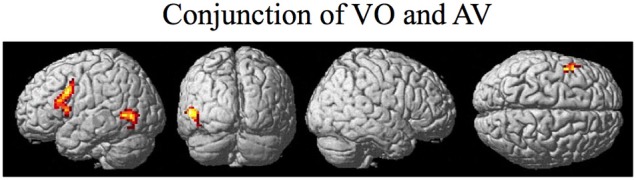
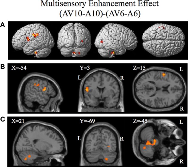
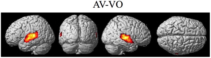
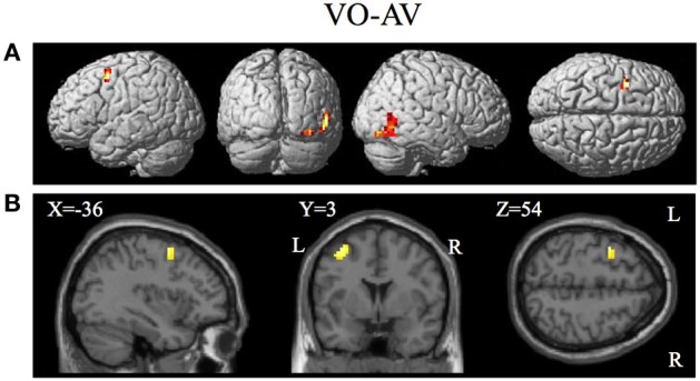
Behavioral and neuroimaging studies have demonstrated that brain regions involved with speech production also support speech perception, especially under degraded conditions. The premotor cortex (PMC) has been shown to be active during both observation and execution of action ("Mirror System" properties), and may facilitate speech perception by mapping unimodal and multimodal sensory features onto articulatory speech gestures. For this functional magnetic resonance imaging (fMRI) study, participants identified vowels produced by a speaker in audio-visual (saw the speaker's articulating face and heard her voice), visual only (only saw the speaker's articulating face), and audio only (only heard the speaker's voice) conditions with varying audio signal-to-noise ratios in order to determine the regions of the PMC involved with multisensory and modality specific processing of visual speech gestures. The task was designed so that identification could be made with a high level of accuracy from visual only stimuli to control for task difficulty and differences in intelligibility. The results of the functional magnetic resonance imaging (fMRI) analysis for visual only and audio-visual conditions showed overlapping activity in inferior frontal gyrus and PMC. The left ventral inferior premotor cortex (PMvi) showed properties of multimodal (audio-visual) enhancement with a degraded auditory signal. The left inferior parietal lobule and right cerebellum also showed these properties. The left ventral superior and dorsal premotor cortex (PMvs/PMd) did not show this multisensory enhancement effect, but there was greater activity for the visual only over audio-visual conditions in these areas. The results suggest that the inferior regions of the ventral premotor cortex are involved with integrating multisensory information, whereas, more superior and dorsal regions of the PMC are involved with mapping unimodal (in this case visual) sensory features of the speech signal with articulatory speech gestures.

Keywords: audio-visual; fMRI; internal model; mirror system; multisensory; premotor.


Figures

Figure 1
Figure 1
Behavioral results as measured by percent correct on the two-alternative forced choice vowel identification task for the following conditions. Audio Visual AV6 (−6 dB SNR), Audio A6 (−6 dB SNR), AV10, A10, AV14, A14, All AV conditions combined, all A conditions combined, Video with noise VN, and Video only VO without noise. All contrasts were significantly greater than chance performance of 50% (p < 0.01).
Figure 2
Figure 2
Behavioral results showing the interaction of audio-visual enhancement at each of the signal-to-noise ratios SNRs. The interaction of (AV6-A6)-(AV10-A10) was statistically significant [F(1, 15) = 12.6, p < 0.005]; however the interaction of (AV10-A10)-(AV14-A14) was not significant [F(1, 15) = 3.9, p > 0.05].
Figure 3
Figure 3
Significant brain activity for the VO condition thresholded at pFDR < 0.05 corrected. Activity was present in the left PMvi/Broca's, left PMvs/PMd, and left and right MT/V5 visual motion processing area.
Figure 4
Figure 4
Significant brain activity for the combined AV conditions thresholded at pFDR < 0.05 corrected. Activity was present in left and right PMvi/Broca's area, left PMvs/PMd, left and right STG/S including primary and secondary auditory cortex, left MT/V5 visual motion processing area, and the right cerebellum lobule VIIb.
Figure 5
Figure 5
Brain activity that was significant for both (conjunction) the VO and the combined AV conditions thresholded at pFDR < 0.05 corrected. Activity was present in the left PMvi/Broca's, left PMvs/PMd, and left MT/V5 visual motion processing area.
Figure 6
Figure 6
Significant brain activity for the contrast that investigated the multisensory enhancement effect (AV10-A10)-(AV6-A6) thresholded at p < 0.001 uncorrected. Activity was present in left PMvi/Broca's area, left pre- and post-central gyrus, left inferior parietal cortex and suprmarginal gyrus, the right occipital lobe, the right cerebellum lobule VIIb and IX, and the left and right brain stem. (A) Activity rendered on the surface of the left, back, right, and top of the brain. (B) Section through brain taken at MNI coordinate −54, 3, 15 shows activity that was present in the PMvi and Broca's region. (C) Section through brain taken at MNI coordinate 21, −69, −45 shows activity that was present in cerebellum lobule VIIb. L, left side of brain; R, right side of brain.
Figure 7
Figure 7
Significant brain activity for the contrast of the combined AV conditions relative to the visual only VO condition thresholded at pFDR < 0.05 corrected. Activity was present in the left and right superior temporal gyrus/sulcus including primary and secondary auditory cortex.
Figure 8
Figure 8
Brain activity significantly active for the contrast of visual only VO relative to the combined AV conditions thresholded at p < 0.001 uncorrected. Activity was present in the left PMvs/PMd and the left MT/V5 visual motion processing area. (A) Activity rendered on the surface of the left, back, right, and top of the brain. (B) Section through brain taken at MNI coordinate −36, 3, 54 shows activity that was present in the PMvs/PMd region. L, left side of brain; R, right side of brain.

References

    1. Alho J., Sato M., Sams M., Schwartz J., Tiitinen H., Jaaskelainen I. (2012). Enhanced early-latency electromagnetic activity in the left premotor cortex is associated with successful phonetic categorization. Neuroimage 60, 1937–1946 10.1016/j.neuroimage.2012.02.011 - DOI - PubMed
    1. Bernstein L., Auer E., Moore J., Ponton C., Don M., Singh M. (2002). Visual speech perception without primary auditory cortex activation. Neuroreport 13, 311–315 10.1097/00001756-200203040-00013 - DOI - PubMed
    1. Callan A., Callan D., Tajima K., Akahane-Yamada R. (2006a). Neural processes involved with perception of non-native durational contrasts. Neuroreport 17, 1353–1357 10.1097/01.wnr.0000224774.66904.29 - DOI - PubMed
    1. Callan D., Callan A., Gamez M., Sato M., Kawato M. (2010). Premotor cortex mediates perceptual performance. Neuroimage 51, 844–858 10.1016/j.neuroimage.2010.02.027 - DOI - PubMed
    1. Callan D., Callan A., Honda K., Masaki S. (2000a). Single-sweep EEG analysis of neural processes underlying perception and production of vowels. Cogn. Brain Res. 10, 173–176 10.1016/S0926-6410(00)00025-2 - DOI - PubMed

LinkOut - more resources