. 2014 May 6:2:e377.
doi: 10.7717/peerj.377. eCollection 2014.

Comparison of histomorphology and DNA preservation produced by fixatives in the veterinary diagnostic laboratory setting

Affiliations

Comparison of histomorphology and DNA preservation produced by fixatives in the veterinary diagnostic laboratory setting

William F Craft et al. PeerJ. .

Abstract

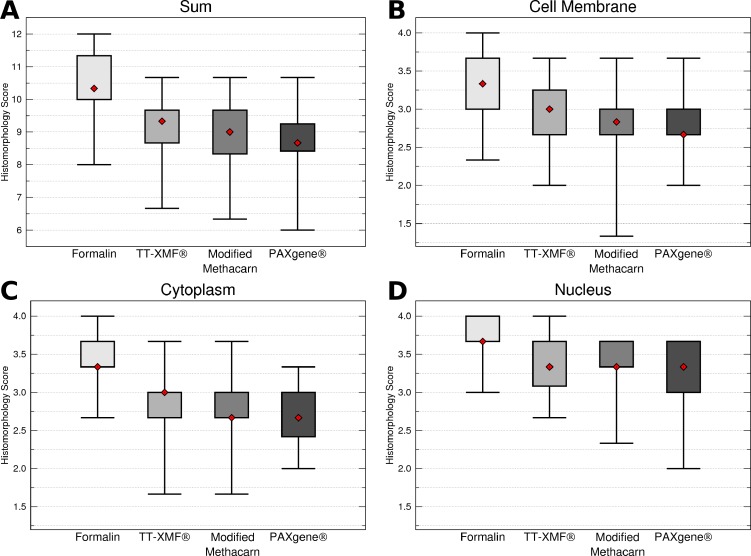
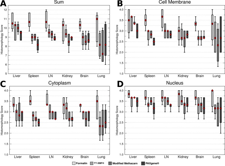
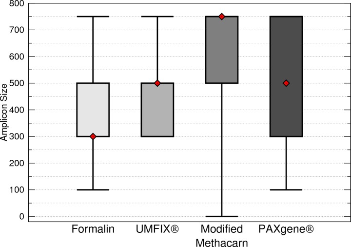
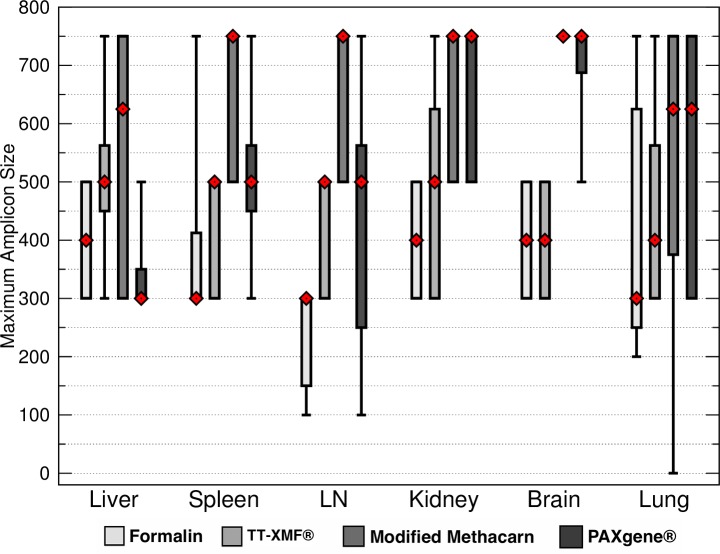
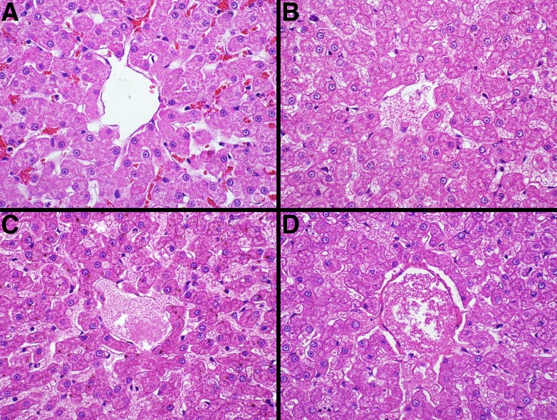
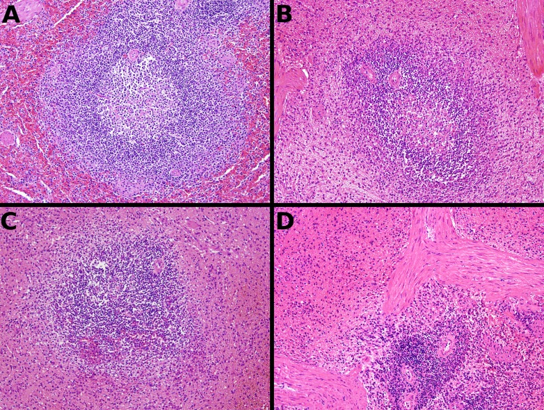
Histopathology is the most useful tool for diagnosis of a number of diseases, especially cancer. To be effective, histopathology requires that tissues be fixed prior to processing. Formalin is currently the most common histologic fixative, offering many advantages: it is cheap, readily available, and pathologists are routinely trained to examine tissues fixed in formalin. However, formalin fixation substantially degrades tissue DNA, hindering subsequent use in diagnostics and research. We therefore evaluated three alternative fixatives, TissueTek(®) Xpress(®) Molecular Fixative, modified methacarn, and PAXgene(®), all of which have been proposed as formalin alternatives, to determine their suitability for routine use in a veterinary diagnostic laboratory. This was accomplished by examining the histomorphology of sections produced from fixed tissues as well as the ability to amplify fragments from extracted DNA. Tissues were sampled from two dogs and four cats, fixed for 24-48 h, and processed routinely. While all fixatives produced acceptable histomorphology, formalin had significantly better morphologic characteristics than the other three fixatives. Alternative fixatives generally had better DNA amplification than formalin, although results varied somewhat depending on the tissue examined. While no fixative is yet ready to replace formalin, the alternative fixatives examined may be useful as adjuncts to formalin in diagnostic practices.

Keywords: Fixative; Formalin; Histomorphology; PCR; Veterinary.


Figures

Figure 1
Figure 1. Comparison of histomorphology and DNA preservation produced by fixatives in the veterinary diagnostic laboratory setting histomorphology scores for all animals and tissues combined.
The median is represented by a red diamond, the box represents the 25th and 75th quartiles, and the whiskers represent 1.5× interquartile range.
Figure 2
Figure 2. Comparison of histomorphology and DNA preservation produced by fixatives in the veterinary diagnostic laboratory setting histomorphology scores for individual tissues.
The median is represented by a red diamond, the box represents the 25th and 75th quartiles, and the whiskers represent 1.5× interquartile range.
Figure 3
Figure 3. Maximum DNA amplicon size ranges for all samples combined.
The median is represented by a red diamond, the box represents the 25th and 75th quartiles, and the whiskers represent 1.5 × interquartile range.
Figure 4
Figure 4. Maximum DNA amplicon size ranges for different tissue samples.
The median is represented by a red diamond, the box represents the 25th and 75th quartiles, and the whiskers represent 1.5 × interquartile range.
Figure 5
Figure 5. Representative fixative histomorphology—liver.
Samples are from the liver of a single cat. A—formalin, B—TT-XMF®, C—modified methacarn, D—PAXgene®.
Figure 6
Figure 6. Representative fixative histomorphology—spleen.
Samples are from the spleen of a single dog. A—formalin, B—TT-XMF®, C—modified methacarn, D—PAXgene®.

References

    1. Bolt HM, Degen GH, Hengstler JG. The carcinogenicity debate on formaldehyde: how to derive safe exposure limits? Archives of Toxicology. 2010;84:421–422. doi: 10.1007/s00204-010-0561-5. - DOI - PubMed
    1. Bosetti C, McLaughlin JK, Tarone RE, Pira E, La Vecchia C. Formaldehyde and cancer risk: a quantitative review of cohort studies through 2006. Annals of Oncology. 2008;19:29–43. doi: 10.1093/annonc/mdm202. - DOI - PubMed
    1. Buesa RJ. Histology without formalin? Annals of Diagnostic Pathology. 2008;12:387–396. doi: 10.1016/j.anndiagpath.2008.07.004. - DOI - PubMed
    1. Cox ML, Schray CL, Luster CN, Stewart ZS, Korytko PJ, Khan KNM, Paulauskis JD, Dunstan RW. Assessment of fixatives, fixation, and tissue processing on morphology and RNA integrity. Experimental and Molecular Pathology. 2006;80:183–191. doi: 10.1016/j.yexmp.2005.10.002. - DOI - PubMed
    1. Fox CH, Johnson FB, Whiting J, Roller PP. Formaldehyde fixation. The Journal of Histochemistry and Cytochemistry. 1985;33:845–853. doi: 10.1177/33.8.3894502. - DOI - PubMed

LinkOut - more resources