. 2014 Feb 1;2(1):66-76.
doi: 10.1007/s40336-014-0051-5.

The Theranostic Path to Personalized Nanomedicine

Affiliations

The Theranostic Path to Personalized Nanomedicine

Benjamin Theek et al. Clin Transl Imaging. .

Abstract

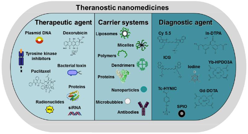
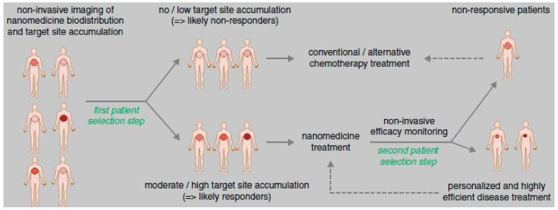
Advances in nanotechnology and chemical engineering have led to the development of many different drug delivery systems. These 1-100(0) nm-sized carrier materials aim to increase drug concentrations at the pathological site, while avoiding their accumulation in healthy non-target tissues, thereby improving the balance between the efficacy and the toxicity of systemic (chemo-) therapeutic interventions. An important advantage of such nanocarrier materials is the ease of incorporating both diagnostic and therapeutic entities within a single formulation, enabling them to be used for theranostic purposes. We here describe the basic principles of using nanomaterials for targeting therapeutic and diagnostic agents to pathological sites, and we discuss how nanotheranostics and image-guided drug delivery can be used to personalize nanomedicine treatments.

Keywords: Drug targeting; Image-guided drug delivery; Nanomedicine; Personalized medicine; Theranostics.


Figures

Figure 1
Figure 1
Schematic representation of carrier materials, therapeutic agents and imaging probes routinely used to prepare theranostic nanomedicine formulations.
Figure 2
Figure 2
Schematic overview of imaging levels in case of anatomical, functional and molecular imaging (A). Anatomical imaging of a fractured mouse femur (B). DCE-MRI of a non-treated or DC101-treated squamous cell carcinoma xenograft four days after therapy start (C). Molecular US imaging of control, RGD and VEGFR2 -coated microbubbles, showing specific binding of RGD and VEGFR2 microbubbles to angiogenic tumor blood vessels (D). Figure adapted from [45,46,37,43].
Figure 3
Figure 3
Nanoparticles as imaging agents. T2-weighted images of liver tumors before (A) and after (B) the i.v. injection of SPIO nanoparticles (tumor indicated by T, spleen by S). Strong negative contrast delineates the exact margins of the tumor. T2-weighted images of LPS-stimulated (C) or native (D) rat brains after the intra-arterial injection of SPIO-labeled VLA-4 expressing human glial precursor cells. Only in the stimulated rat (C), a negative contrast, corresponding to cell retention, is observed. MR images obtained 35 min after the i.v. injection of RGD-conjugated (E) vs. RAD-modified control liposomes (F) into tumor-bearing mice. The respective fluorescence images of the tumor are shown in (G) and (H). Figures adapted from [50,52,54].
Figure 4
Figure 4
Image-guided drug delivery. 2D fluorescence reflectance image (FRI; A) and 3D fluorescence molecular tomography (FMT; B) of a near-infrared fluorophore-labeled HPMA copolymer in mice bearing subcutaneous CT26 tumors. Gamma-scintigraphy of patients 24 h after the i.v. injection of radiolabeled PK1 (pHPMA-doxorubicin; left) and PK2 (galactosamine-targeted pHPMA-doxorubicin; right) (C). Gamma-camera images obtained at 72 h after the i.v. injection of indium-labeled PEGylated liposomes in breast cancer (D), lung cancer (E), and head-and-neck cancer (F). Images demonstrate accumulation in tumor (T), lymph node (LN), liver (L), spleen (S) and cardiac blood pool (CP). Figures adaped from [55,57,56].
Figure 5
Figure 5
Nanotheranostics for personalized nanomedicine. Patients to be treated with nanomedicines are to be prescreened prior to therapy with a (radio-) labeled version of the formulation (first patient selection step), in order to identify individuals showing sufficiently high levels of (EPR-mediated) tumor accumulation, and which therefore are more likely to respond to treatment with the targeted nanomedicine formulation in question. During a second patient selection step, individuals showing reasonable target site accumulation but insufficient therapeutic response could be allocated to treatment with alternative therapies, to assure individualized and improved interventions. Figure adapted from [60].

References

    1. Hanahan D, Weinberg RA. Hallmarks of cancer: the next generation. Cell. 2011;144(5):646–674. - PubMed
    1. Hanahan D, Weinberg RA. The hallmarks of cancer. Cell. 2000;100(1):57–70. - PubMed
    1. Essenberg JM. Cigarette smoke and the incidence of primary neoplasm of the lung in the albino mouse. Science. 1952;116(3021):561–562. - PubMed
    1. Hecht SS. Lung carcinogenesis by tobacco smoke. Int J Cancer. 2012;131(12):2724–2732. - PMC - PubMed
    1. Fearon ER. Molecular genetics of colorectal cancer. Annu Rev Pathol. 2011;6:479–507. - PubMed

LinkOut - more resources