

. 2014 Jul;42(Web Server issue):W215-20.
doi: 10.1093/nar/gku460.
Epub 2014 May 26.
ProBiS-ligands: a web server for prediction of ligands by examination of protein binding sites
Affiliations
- PMID: 24861616
- PMCID: PMC4086080
- DOI: 10.1093/nar/gku460
Item in Clipboard
ProBiS-ligands: a web server for prediction of ligands by examination of protein binding sites
Nucleic Acids Res.
2014 Jul.
Abstract
The ProBiS-ligands web server predicts binding of ligands to a protein structure. Starting with a protein structure or binding site, ProBiS-ligands first identifies template proteins in the Protein Data Bank that share similar binding sites. Based on the superimpositions of the query protein and the similar binding sites found, the server then transposes the ligand structures from those sites to the query protein. Such ligand prediction supports many activities, e.g. drug repurposing. The ProBiS-ligands web server, an extension of the ProBiS web server, is open and free to all users at http://probis.cmm.ki.si/ligands.
© The Author(s) 2014. Published by Oxford University Press on behalf of Nucleic Acids Research.
Figures
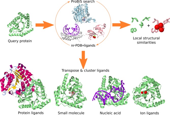
Ligand prediction by the ProBiS-ligands server, starting from a query protein structure (light green).
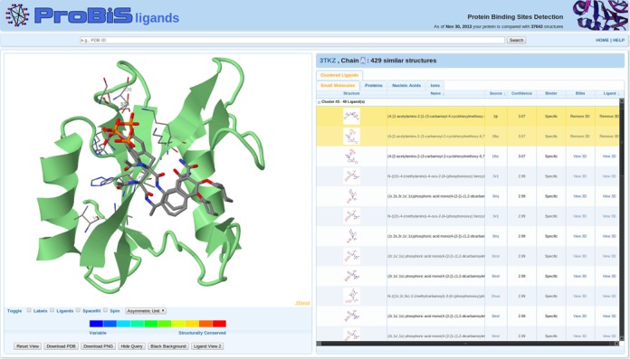
ProBiS-ligands output page. Left: query protein (green cartoon model) and two predicted ligands (CPK colored stick models). Invariant binding sites residues are thinner CPK-colored sticks. Right: table with predicted small-molecule ligands clustered according to their predicted location on the query protein and transposed from different binding sites; the two selected ligands are highlighted.
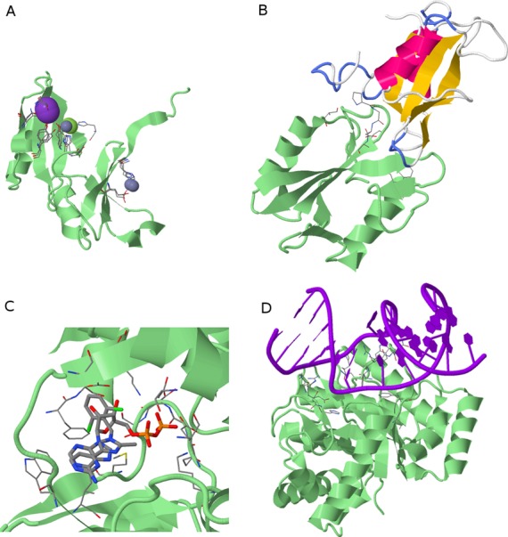
Predicted protein–ligand complexes. Query proteins are green cartoon models and invariant binding site residues are CPK-colored stick models (Ligand View 1). (A) Three predicted ion ligand clusters (ions are spheres) on Glyoxalase family protein (PDB ID: 2qqz). (B) Predicted protein ligand (yellow–pink cartoon) on SH2 domain protein (3tkz). (C) Two predicted small molecule ligands, i.e. ATP and an inhibitor of biotin carboxylase (thick CPK sticks) on D-alanine:D-alanine ligase (1iov). (D) Predicted DNA ligand on endonuclease IV protein (4hno).
References
-
- Yuriev E., Ramsland P.A. Latest developments in molecular docking: 2010–2011 in review. J. Mol. Recognit. 2013;26:215–239. - PubMed
-
- Haupt V.J., Schroeder M. Old friends in new guise: repositioning of known drugs with structural bioinformatics. Briefings Bioinform. 2011;12:312–326. - PubMed
-
- Moriaud F., Richard S.B., Adcock S.A., Chanas-Martin L., Surgand J.-S., Jelloul M.B., Delfaud F. Identify drug repurposing candidates by mining the Protein Data Bank. Briefings Bioinform. 2011;12:336–340. - PubMed
Publication types
MeSH terms
Substances
Associated data
- Actions
- Actions
- Actions
- Actions
- Actions
LinkOut - more resources
Full Text Sources
Other Literature Sources
