. 2014 Jul;42(13):e104.
doi: 10.1093/nar/gku439. Epub 2014 May 26.

Quantitative telomerase enzyme activity determination using droplet digital PCR with single cell resolution

Affiliations

Quantitative telomerase enzyme activity determination using droplet digital PCR with single cell resolution

Andrew T Ludlow et al. Nucleic Acids Res. 2014 Jul.

Abstract

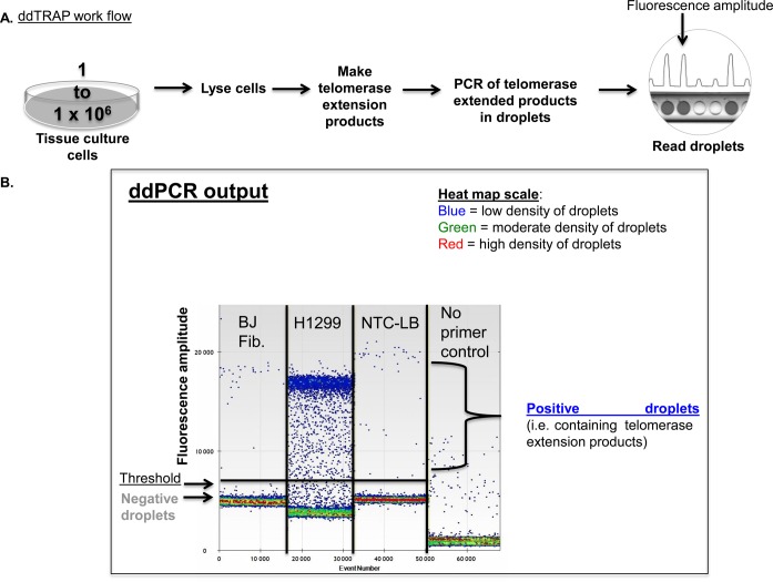
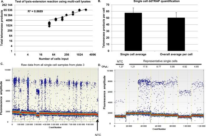
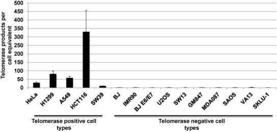
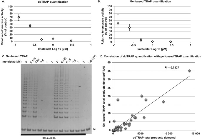
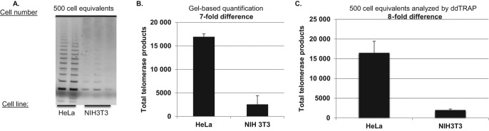
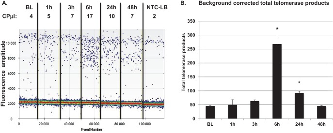
The telomere repeat amplification protocol (TRAP) for the human reverse transcriptase, telomerase, is a PCR-based assay developed two decades ago and is still used for routine determination of telomerase activity. The TRAP assay can only reproducibly detect ∼ 2-fold differences and is only quantitative when compared to internal standards and reference cell lines. The method generally involves laborious radioactive gel electrophoresis and is not conducive to high-throughput analyzes. Recently droplet digital PCR (ddPCR) technologies have become available that allow for absolute quantification of input deoxyribonucleic acid molecules following PCR. We describe the reproducibility and provide several examples of a droplet digital TRAP (ddTRAP) assay for telomerase activity, including quantitation of telomerase activity in single cells, telomerase activity across several common telomerase positive cancer cells lines and in human primary peripheral blood mononuclear cells following mitogen stimulation. Adaptation of the TRAP assay to digital format allows accurate and reproducible quantification of the number of telomerase-extended products (i.e. telomerase activity; 57.8 ± 7.5) in a single HeLa cell. The tools developed in this study allow changes in telomerase enzyme activity to be monitored on a single cell basis and may have utility in designing novel therapeutic approaches that target telomerase.


Figures

Figure 1.
Figure 1.
Workflow and optimization of droplet digital TRAP. (A). The ddTRAP workflow. Cells are lysed at a concentration of 1250 cells per μl, telomerase extension products generated at a concentration of 25 cells/μl, then telomerase is heat inactivated and extension products dispersed into droplets. PCR thermocycling is done for 40 cycles and droplets analyzed for the presence or absence of fluorescence by the droplet reader. (B). ddTRAP output showing BJ fibroblasts (input of 100 cell equivalents, telomerase negative), H1299 cells (input of 100 cell equivalents, telomerase positive), a lysis buffer only control and a control with no primers and input of 100 cell equivalents of H1299 lysate to test for specificity of amplification. Only very low background signals are seen in these controls. Each well or sample of the ddPCR analyzes about 17 000 droplets. Event number at the bottom of the output represents the number of droplets counted in the wells overtime. Each dot on the ddPCR output represents a unique droplet that is either positive or negative for fluorescent signal. Fluorescence amplitude is a measure of the fluorescence detected for each droplet in the assay. Fluorescence amplitude is used to separate the positive and negative droplets. Since the droplets are detected with EvaGreen® double stranded DNA binding dye there will be inherent background fluorescence of DNA molecules not amplified during PCR. The heat map scale represents the density of droplets at given fluorescent amplitudes. NTC-LB = no-template control-lysis buffer.
Figure 2.
Figure 2.
Optimization of ddTRAP. (A) ddTRAP fluorescent droplet outputs are shown on the right, and quantified outputs graphed on the left. Lysates were incubated with TS substrate for various amounts of time prior to heat inactivation, using 100 cell equivalents from H1299 cells. Data are presented as means of background corrected total telomerase products generated ± standard error of the mean. Background corrected total telomerase products generated was calculated by first multiplying the concentration of molecules counted (molecules per microliter) by 20 (for the 20 μl PCR) and then subtracting the mean of the NTC-LB control from each sample. (B) Heat and RNase A inactivation of telomerase resulted in virtually no ddTRAP signal. This permitted a small background correction to be included, and established the specific measurement of telomerase activity from lysates. Background signal is highlighted by the red box.
Figure 3.
Figure 3.
Reproducibility and linearity of ddTRAP. (A) Intra-day variability was assessed by making three extension reaction dilution series and assaying telomerase activity on one plate for each cell input (three replicates per-cell equivalent input). A clone of the HeLa cell line specific to our lab observed to have intermediate telomerase activity (was used in this experiment see Figures 4 and 6 for HeLa cell population telomerase activity). Data are plotted as total telomerase product (copies of target molecules per microliter multiplied by 20 = copies per μl) by cell equivalents. Error bars are standard deviation of the replicates on each plate. (B) Inter-day variability was assessed by running two assays on two separate days from the same three extension reactions described in (A). Data are plotted as total telomerase product (copies per microliter multiplied by 20) by cell equivalents. Error bars are standard deviation of the replicates on each plate. (C) Technical variation was assayed by diluting the 100 cell equivalent sample 1:2 in a series of six samples. Telomerase activity was measured on two separate plates on the same day. Technical variation is plotted in copies per microliter (output from ddPCR software) by cell equivalents input on a log scale. Error bars are Poisson corrected 95% confidence intervals across the two plates. (D) HeLa cell dilution series from 50 to 1 cell equivalents produced a linear relationship (R2 = 0.99) between input and detection of telomerase enzyme activity as indicated by total product generated in the ddPCR. Error bars are the standard deviation of the replicates. Although the extract was not made from a single cell, ddTRAP was able to reproducibly detect telomerase activity above background at the dilution equivalent to one cell input.
Figure 4.
Figure 4.
Telomerase activity in single cells. Telomerase activity (i.e. the number of extended TS molecules counted following ddPCR) in single cells was determined using a lysis and extension reaction. (A) The lysis-extension buffer is linear and produced similar results to the two-step lysis-extension procedure previously used. The slightly higher values are likely due to more efficient lysis using this new buffer system. Note that the scale of the graph is log(4). (B) Telomerase activity from single HeLa cells. 2 μl of two dilutions of cells (3000 cells/μl and 300 cells/μl) were plated overnight in 6-well plates to allow single cells to be visualized with a light microscope, then dissolved in 2 μl of lysis-extension mixture. We picked both single cells (n = 78) and groups of 2–8 cells. The 2 μl lysis-extension reaction was incubated on ice for at least 30 min, then placed in a thermocycler for the telomerase extension reaction at 25ºC for 40 min. Following extension, 18 μl of ddPCR mixture (10 μl 2× EvaGreen ddPCR Supermix®, 0.1 μl of 10 μM TS primer, 0.1 μl of 10 μM ACX primer and 7.8 μl of dH2O) was added to the 2 μl lysis-extension reaction, droplets were produced, PCR performed and fluorescence of the droplets read. The ddPCR in copies per microliter was corrected for background (subtraction of the copies per microliter value of the no-template lysis buffer control). The corrected copies per microliter values were then multiplied by 20 to give the total telomerase products values. An average of 57.8 telomerase-extended molecules were observed per-cell. Telomerase activity assayed was similar in single cell lysates compared to telomerase activity detected in lysates generated from greater numbers of cells using the lysis-extension buffer and normalized to a per-cell equivalent average (range from 2 to 8 cells). (C) Raw ddPCR output of all single cell lanes from the third 96-well plate that was run using the lysis extension system. (D) Representative lanes of ddPCR output showing the NTC-LB control, a negative sample and several samples of various telomerase activities. CPμl = copies of target per microliter, output from the droplet reader software.
Figure 5.
Figure 5.
Telomerase activity across different cell types. Cancer cell lines known to be telomerase positive gave ddTRAP signals while cell lines known to be telomerase negative gave no signal following background correction. 50 cell equivalents from each cell line were analyzed with ddTRAP. Telomerase activity is defined as the number of extended TS molecules counted following ddPCR and then multiplied by 20 and divided by the number of cell equivalents added into the PCR to produce normalized activity values.
Figure 6.
Figure 6.
Comparison of ddTRAP to gel based TRAP: determination of Imetelstat IC50 in HeLa cells. ddTRAP quantification is less variable than gel based TRAP and allowed accurate determination of the IC50 of Imetelstat in HeLa (0.2 μM). HeLa cells were incubated with Imetelstat (0, 0.125, 0.25, 0.5, 1 and 3 μM) for 72 h. Cells were pelleted and triplicate extracts prepared from three separate tissue culture experiments (nine total extracts and extensions per dose). (A) ddTRAP quantification with 50 cell equivalents added to the PCR. (B) Gel quantification (representative gel image of two experiments in Figure 5C). (C) Gel based TRAP was performed with 125 cell equivalents. Data are expressed as relative telomerase activity compared to control (untreated HeLa) and standard error of the mean. (D) Correlation analysis of gel-based TRAP to ddTRAP. The P < 0.0001 positive relationship indicates that the methods are measuring the same phenomenon. ITAS = Internal competitive telomerase activity substrate.
Figure 7.
Figure 7.
ddTRAP detection of mouse telomerase. Extracts from one million HeLa and one million NIH3T3 mouse fibroblasts were prepared. Following lysis (40 μl), 1-μl of the lysate (25 000 cell/μl) was added to a 50 μl extension reaction (extension product concentration = 500 cells/μl). One-microliter of the extension reaction was used for either the gel based TRAP or the ddTRAP assay. (A) Gel analysis (10% PAGE) of HeLa compared to NIH3T3 cells. Gel was stained with Gel red, a double stranded DNA binding dye. (B) ddTRAP total products generated (molecules per microliter output multiplied by 20 μl). Quantification showed an eight-fold difference in extension products Eqs. = equivalents. (C) Quantification of scanned images from A (ImageJ, http://imagej.nih.gov/ij/, (22)) showing a seven-fold difference which is similar to ddTRAP quantification.
Figure 8.
Figure 8.
ddTRAP detects mitogen-stimulated telomerase in human PBMCs. Fifty cell equivalents were assayed using ddTRAP. (A) Raw ddPCR output. Telomerase activity was detected above background at 6 h of PHA stimulation that persisted until 24 h and returned to baseline levels at 48 h of stimulation. (B) Calculated total telomerase products generated during the extension reaction (ddPCR output in copies per microliter multiplied by 20). Data are background corrected. CPμl = copies of target per microliter, output from the droplet reader software. *Significantly different from control P < 0.05 by Student's Paired t-test.

Similar articles

Cited by

References

    1. Daniali L., Benetos A., Susser E., Kark J.D., Labat C., Kimura M., Desai K., Granick M., Aviv A. Telomeres shorten at equivalent rates in somatic tissues of adults. Nat. Commun. 2013;4:1597–1600. - PMC - PubMed
    1. Shay J.W., Wright W.E. Telomeres and telomerase in normal and cancer stem cells. FEBS Lett. 2010;584:3819–3825. - PMC - PubMed
    1. Kim N.W., Piatyszek M.A., Prowse K.R., Harley C.B., West M.D., Ho P.L., Coviello G.M., Wright W.E., Weinrich S.L., Shay J.W. Specific association of human telomerase activity with immortal cells and cancer. Science. 1994;266:2011–2015. - PubMed
    1. Harley C.B., Kim N.W., Prowse K.R., Weinrich S.L., Hirsch K.S., West M.D., Bacchetti S., Hirte H.W., Counter C.M., Greider C.W., et al. Telomerase, cell immortality, and cancer. Cold Spring Harb. Symp. Quant. Biol. 1994;59:307–315. - PubMed
    1. Greider C.W. Mammalian telomere dynamics: healing, fragmentation shortening and stabilization. Curr. Opin. Genet. Dev. 1994;4:203–211. - PubMed

Publication types