

Measurement of ADP-ATP exchange in relation to mitochondrial transmembrane potential and oxygen consumption
- PMID: 24862274
- PMCID: PMC4630003
- DOI: 10.1016/B978-0-12-416618-9.00017-0
Measurement of ADP-ATP exchange in relation to mitochondrial transmembrane potential and oxygen consumption
Abstract
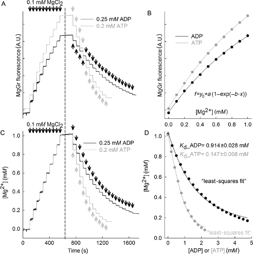
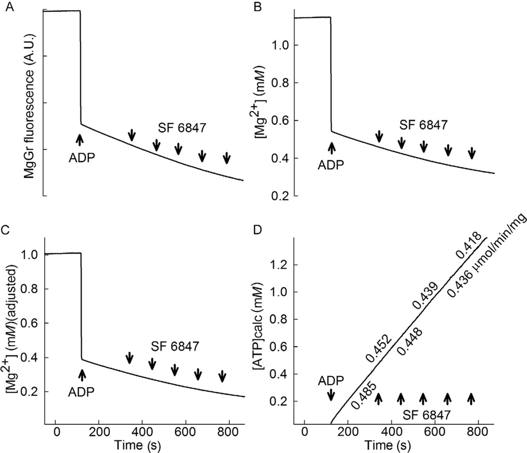
We have previously described a fluorometric method to measure ADP-ATP exchange rates in mitochondria of permeabilized cells, in which several enzymes that consume substantial amounts of ATP and other competing reactions interconverting adenine nucleotides are present. This method relies on recording changes in free extramitochondrial Mg(2+) with the Mg(2+)-sensitive fluorescent indicator Magnesium Green (MgGr)™, exploiting the differential affinity of ADP and ATP for Mg(2+). In particular, cells are permeabilized with digitonin in the presence of BeF3(-) and Na3VO4, inhibiting all ATP- and ADP-utilizing reactions but mitochondrial exchange of ATP with ADP catalyzed by the adenine nucleotide translocase. The rate of ATP appearing in the medium upon the addition of ADP to energized mitochondria is then calculated from the rate of change in free extramitochondrial Mg(2+) using standard binding equations. Here, we describe a variant of this method involving an improved calibration step. This step minimizes errors that may be introduced during the conversion of the MgGr™ signal into free extramitochondrial [Mg(2+)] and ATP. Furthermore, we describe an approach for combining this methodology with the measurement of mitochondrial membrane potential and oxygen consumption in the same sample. The method described herein is useful for the study of malignant cells, which are known to thrive in hypoxic environments and to harbor mitochondria with profound functional alterations.
Keywords: ADP–ATP exchange; Adenine nucleotide carrier; Adenine nucleotide translocase; Bioenergetics; Oxidative phosphorylation; Systems biology.
© 2014 Elsevier Inc. All rights reserved.
Figures

References
-
- Akerman KE, Wikstrom MK. Safranine as a probe of the mitochondrial membrane potential. FEBS Letters. 1976;68:191–197. - PubMed
-
- Aprille JR. Mechanism and regulation of the mitochondrial ATP-Mg/P(i) carrier. Journal of Bioenergetics and Biomembranes. 1993;25:473–481. - PubMed
-
- Aureliano M, Crans DC. Decavanadate (V10 O28 6-) and oxovanadates: Oxometalates with many biological activities. Journal of Inorganic Biochemistry. 2009;103:536–546. - PubMed
-
- Baukrowitz T, Hwang TC, Nairn AC, Gadsby DC. Coupling of CFTR Cl- channel gating to an ATP hydrolysis cycle. Neuron. 1994;12:473–482. - PubMed
-
- Byczkowski J, Zychlinski L, Tluczkiewicz J. Interaction of vanadate with respiratory chain of rat liver and wheat seedling mitochondria. The International Journal of Biochemistry. 1979;10:1007–1011. - PubMed
Publication types
MeSH terms
Substances
Grants and funding
LinkOut - more resources
Full Text Sources
Other Literature Sources
