Review
. 2014 Jul 15;426(14):2539-46.
doi: 10.1016/j.jmb.2014.05.020. Epub 2014 May 24.

Competition as a way of life for H(+)-coupled antiporters

Affiliations
Review

Competition as a way of life for H(+)-coupled antiporters

Shimon Schuldiner. J Mol Biol. .

Abstract

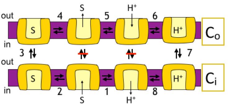
Antiporters are ubiquitous membrane proteins that catalyze obligatory exchange between two or more substrates across a membrane in opposite directions. Some utilize proton electrochemical gradients generated by primary pumps by coupling the downhill movement of one or more protons to the movement of a substrate. Since the direction of the proton gradient usually favors proton movement toward the cytoplasm, their function results in removal of substrates other than protons from the cytoplasm, either into acidic intracellular compartments or out to the medium. H(+)-coupled antiporters play central roles in living organisms, for example, storage of neurotransmitter and other small molecules, resistance to antibiotics, homeostasis of ionic content and more. Biochemical and structural data support a general mechanism for H(+)-coupled antiporters whereby the substrate and the protons cannot bind simultaneously to the protein. In several cases, it was shown that the binding sites overlap, and therefore, there is a direct competition between the protons and the substrate. In others, the "competition" seems to be indirect and it is most likely achieved by allosteric mechanisms. The pKa of one or more carboxyls in the protein must be tuned appropriately in order to ensure the feasibility of such a mechanism. In this review, I discuss in detail the case of EmrE, a multidrug transporter from Escherichia coli and evaluate the information available for other H(+)-coupled antiporters.

Keywords: coupling mechanism; membrane proteins; multidrug transporters; transport; vesicular neurotransmitter transporters.


Figures

Figure 1
Figure 1. A simplified view of the catalytic cycle of an antiporter
For the sake of simplicity the cartoon includes only the conformations where the protein faces the inside (Ci) or the outside (Co) with a proton (CiH and CoH) or substrate (CiS and COS) bound. Modified from figure 1 in [44]
Figure 2
Figure 2. pH dependence of the equilibrium KD of EmrE
Purified protein was immobilized on Ni-NTA beads and bound to increasing [3H]TPP+ concentrations at various pH values as described [9]. Adapted from [9].
Figure 3
Figure 3. Partial steps of the cycle
Rates of substrate binding are dependent on pH because it binds only the unprotonated form of the transporter (bottom panel, x-x). Downhill transport of substrate includes all the steps in the cycle described in figure 1 (top left). The pH on both sides of the membrane is kept the same by the ionophore Nigericin and the reaction is driven by the substrate electrochemical gradient. The pH dependence of the reaction is shown at the bottom (◆-◆). The exchange reaction between radiolabeled (*MV2+) and unlabeled (MV2+) Methyl Viologen (top middle) does not include the proton translocation steps (6-8 in figure 1) but since rates of substrate binding are affected by pH also the exchange reaction shows pH dependence similar to that of the binding reaction and the downhill transport (▲-▲). The dependence of H+-driven uptake (■-■) is shifted about 1 pH unit to the alkaline side. In these experiments, at each external pH the internal pH is about 2 pH units lower. A likely interpretation of this shift is that Glu14 is exposed alternately to the internal and external pH of the proteoliposome and “senses” the average pH between the inside and the outside. Adapted from [14].
Figure 4
Figure 4. Tryptophan fluorescence spectra of EmrE-single Trp63 at different pH values with/without substrate
A- Emission spectra (excitation at 280 nm) of 2 μM EmrE-Single Trp 63. The lowest solid line is at pH 5.5 the middle is at pH 7.0 and the highest is at pH 8.5. The dashed line represents protein at pH 8.5 after addition of 50 μM of the substrate TPP+. The shoulder at around 310 nm in the spectra of the single Trp63 mutant (that bears 7 Tyr residues) is not observed upon excitation at 295 nm and was assigned to Tyr fluorescence. B- Summary of the pH dependence of the fluorescence intensity at the emission peak at 338 nm of free protein (squares) and substrate bound protein (circles). Adapted from [9].

References

    1. Donowitz M, Ming Tse C, Fuster D. SLC9/NHE gene family, a plasma membrane and organellar family of Na+/H+ exchangers. Mol Aspects Med. 2013;34:236–51. - PMC - PubMed
    1. Accardi A, Picollo A. CLC channels and transporters: proteins with borderline personalities. Biochim Biophys Acta. 2010;1798:1457–64. - PMC - PubMed
    1. Lawal HO, Krantz DE. SLC18: Vesicular neurotransmitter transporters for monoamines and acetylcholine. Mol Aspects Med. 2013;34:360–72. - PMC - PubMed
    1. Fluman N, Bibi E. Bacterial multidrug transport through the lens of the major facilitator superfamily. Biochim Biophys Acta. 2009;1794:738–47. - PubMed
    1. Waight AB, Pedersen BP, Schlessinger A, Bonomi M, Chau BH. Roe-Zurz Z, et al. Structural basis for alternating access of a eukaryotic calcium/proton exchanger. Nature. 2013;499:107–10. - PMC - PubMed

Publication types

LinkOut - more resources