

Polymer multilayers loaded with antifungal β-peptides kill planktonic Candida albicans and reduce formation of fungal biofilms on the surfaces of flexible catheter tubes
- PMID: 24862322
- PMCID: PMC4156914
- DOI: 10.1016/j.jconrel.2014.05.026
Polymer multilayers loaded with antifungal β-peptides kill planktonic Candida albicans and reduce formation of fungal biofilms on the surfaces of flexible catheter tubes
Abstract
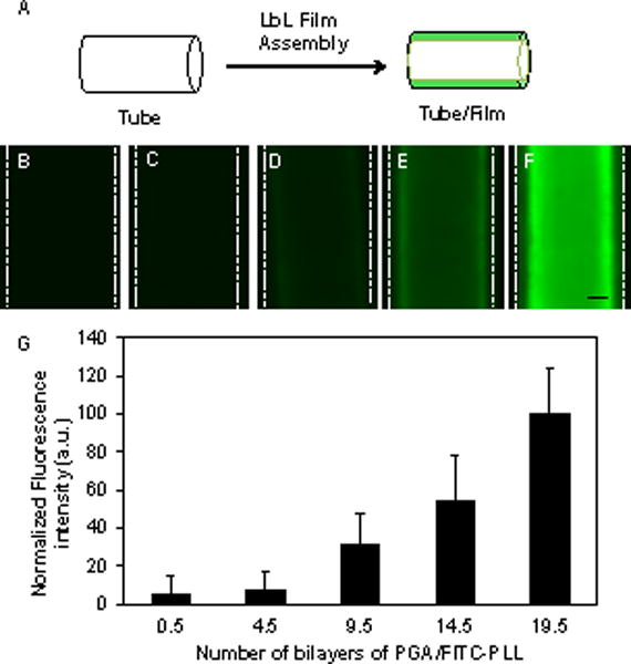
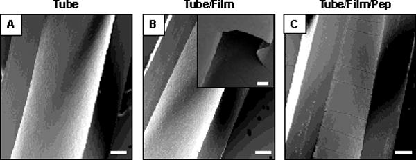
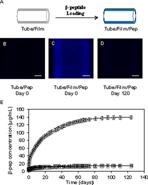
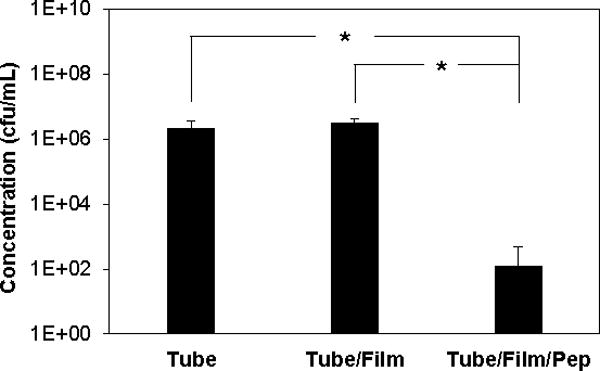
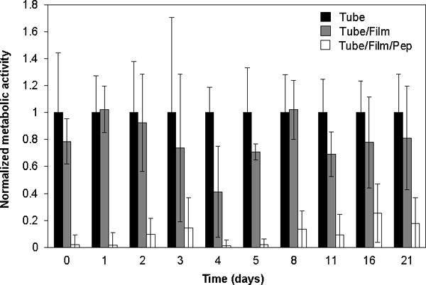
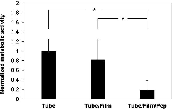
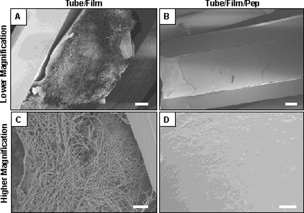
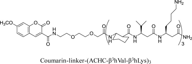
Candida albicans is the most common fungal pathogen responsible for hospital-acquired infections. Most C. albicans infections are associated with the implantation of medical devices that act as points of entry for the pathogen and as substrates for the growth of fungal biofilms that are notoriously difficult to eliminate by systemic administration of conventional antifungal agents. In this study, we report a fill-and-purge approach to the layer-by-layer fabrication of biocompatible, nanoscale 'polyelectrolyte multilayers' (PEMs) on the luminal surfaces of flexible catheters, and an investigation of this platform for the localized, intraluminal release of a cationic β-peptide-based antifungal agent. We demonstrate that polyethylene catheter tubes with luminal surfaces coated with multilayers ~700nm thick fabricated from poly-l-glutamic acid (PGA) and poly-l-lysine (PLL) can be loaded, post-fabrication, by infusion with β-peptide, and that this approach promotes extended intraluminal release of this agent (over ~4months) when incubated in physiological media. The β-peptide remained potent against intraluminal inoculation of the catheters with C. albicans and substantially reduced the formation of C. albicans biofilms on the inner surfaces of film-coated catheters. Finally, we report that these β-peptide-loaded coatings exhibit antifungal activity under conditions that simulate intermittent catheter use and microbial challenge for at least three weeks. We conclude that β-peptide-loaded PEMs offer a novel and promising approach to kill C. albicans and prevent fungal biofilm formation on surfaces, with the potential to substantially reduce the incidence of device-associated infections in indwelling catheters. β-Peptides comprise a promising new class of antifungal agents that could help address problems associated with the use of conventional antifungal agents. The versatility of the layer-by-layer approach used here thus suggests additional opportunities to exploit these new agents in other biomedical and personal care applications in which fungal infections are endemic.
Keywords: Antifungal; Biofilms; Catheters; Controlled release; Polymer multilayers; Surfaces.
Copyright © 2014 Elsevier B.V. All rights reserved.
Figures
Similar articles
-
Polyelectrolyte multilayers fabricated from antifungal β-peptides: design of surfaces that exhibit antifungal activity against Candida albicans.Biomacromolecules. 2010 Sep 13;11(9):2321-8. doi: 10.1021/bm100424s. Biomacromolecules. 2010. PMID: 20831274 Free PMC article.
-
Antifungal activity of a β-peptide in synthetic urine media: Toward materials-based approaches to reducing catheter-associated urinary tract fungal infections.Acta Biomater. 2016 Oct 1;43:240-250. doi: 10.1016/j.actbio.2016.07.016. Epub 2016 Jul 12. Acta Biomater. 2016. PMID: 27422198 Free PMC article.
-
Intraluminal Release of an Antifungal β-Peptide Enhances the Antifungal and Anti-Biofilm Activities of Multilayer-Coated Catheters in a Rat Model of Venous Catheter Infection.ACS Biomater Sci Eng. 2016 Jan 11;2(1):112-121. doi: 10.1021/acsbiomaterials.5b00427. Epub 2015 Dec 8. ACS Biomater Sci Eng. 2016. PMID: 26807439 Free PMC article.
-
Antimicrobial Peptides as a Strategy to Combat Fungal Biofilms.Curr Top Med Chem. 2017;17(5):604-612. doi: 10.2174/1568026616666160713142228. Curr Top Med Chem. 2017. PMID: 27411323 Review.
-
Impact of multiscale surface topography characteristics on Candida albicans biofilm formation: From cell repellence to fungicidal activity.Acta Biomater. 2024 Mar 15;177:20-36. doi: 10.1016/j.actbio.2024.02.006. Epub 2024 Feb 9. Acta Biomater. 2024. PMID: 38342192 Review.
Cited by
-
Holistic Molecular Design of Ionic Surfaces for Tailored Water Wettability and Technical Applications.Nanomaterials (Basel). 2025 Apr 11;15(8):591. doi: 10.3390/nano15080591. Nanomaterials (Basel). 2025. PMID: 40278457 Free PMC article. Review.
-
Streptomyces: Derived Active Extract Inhibits Candida albicans Biofilm Formation.Curr Microbiol. 2022 Sep 25;79(11):332. doi: 10.1007/s00284-022-03013-1. Curr Microbiol. 2022. PMID: 36155861
-
Polyelectrolyte Multilayers on Soft Colloidal Nanosurfaces: A New Life for the Layer-By-Layer Method.Polymers (Basel). 2021 Apr 9;13(8):1221. doi: 10.3390/polym13081221. Polymers (Basel). 2021. PMID: 33918844 Free PMC article. Review.
-
Histatin 5 variant reduces Candida albicans biofilm viability and inhibits biofilm formation.Fungal Genet Biol. 2021 Apr;149:103529. doi: 10.1016/j.fgb.2021.103529. Epub 2021 Feb 14. Fungal Genet Biol. 2021. PMID: 33596477 Free PMC article.
-
Development of antimicrobial coating by layer-by-layer [corrected] dip coating of chlorhexidine-loaded micelles.J Mater Sci Mater Med. 2017 Jun;28(6):90. doi: 10.1007/s10856-017-5899-2. Epub 2017 May 9. J Mater Sci Mater Med. 2017. PMID: 28488039
References
-
- Azie N, Neofytos D, Pfaller M, Meier-Kriesche HU, Quan SP, Horn D. The PATH (Prospective Antifungal Therapy) Alliance® registry and invasive fungal infections: update 2012. Diagn Microbiol Infect Dis. 2012;73:293–300. - PubMed
-
- Gullo A. Invsasive Fungal Infections: The Challenge Continues. Drugs. 2009;69:65–73. - PubMed
-
- Brown GD, Denning DW, Gow NaR, Levitz SM, Netea MG, White TC. Hidden killers: human fungal infections. Sci Transl Med. 2012;4:165rv13. - PubMed
-
- Wenzel RP. Nosocomial Candidemia : Risk Factors and Attributable Mortality. Clin Infect Dis. 1995;20:1531–1534. - PubMed
-
- Calderone Ra, Fonzi Wa. Virulence factors of Candida albicans. Trends Microbiol. 2001;9:327–35. - PubMed
Publication types
MeSH terms
Substances
Grants and funding
LinkOut - more resources
Full Text Sources
Other Literature Sources
