Review
. 2014 May 26;205(4):435-45.
doi: 10.1083/jcb.201402054.

Organellophagy: eliminating cellular building blocks via selective autophagy

Affiliations
Review

Organellophagy: eliminating cellular building blocks via selective autophagy

Koji Okamoto. J Cell Biol. .

Abstract

Maintenance of organellar quality and quantity is critical for cellular homeostasis and adaptation to variable environments. Emerging evidence demonstrates that this kind of control is achieved by selective elimination of organelles via autophagy, termed organellophagy. Organellophagy consists of three key steps: induction, cargo tagging, and sequestration, which involve signaling pathways, organellar landmark molecules, and core autophagy-related proteins, respectively. In addition, posttranslational modifications such as phosphorylation and ubiquitination play important roles in recruiting and tailoring the autophagy machinery to each organelle. The basic principles underlying organellophagy are conserved from yeast to mammals, highlighting its biological relevance in eukaryotic cells.


Figures

Figure 1.
Figure 1.
Three distinct modes of autophagy. In macroautophagy, newly generated cup-shaped structures, called isolation membranes, expand to surround cytoplasmic components. The two edges of isolation membranes then fuse to form double membrane–bound autophagosomes. Subsequently, autophagosomes fuse to lysosomes, and the engulfed cargoes are digested by hydrolytic enzymes. In microautophagy, invagination of the lysosomal membrane occurs to sequester proteins and organelles in the cytosol. The resulting vesicular structures are then pinched off and released into the lysosomal lumen for digestion. In chaperone-mediated autophagy (CMA), the Hsc70/co-chaperone complex delivers specific substrate proteins to lysosomes. The substrate polypeptides are then translocated one by one through the lysosomal membrane protein Lamp-2A and digested in the lysosomal lumen. Macro- and microautophagy are conserved from yeast to humans, whereas CMA has been found only in mammals. Unlike macro- and microautophagy, CMA has been suggested to degrade only proteins but not whole organelles.
Figure 2.
Figure 2.
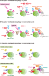
Two common mechanisms of organellophagy. Molecular mechanisms underlying cargo recognition in pexophagy and mitophagy have extensively been explored, including two common types, receptor- and ubiquitin-mediated processes. Both types involve protein phosphorylation that activates or inactivates their downstream events. In the receptor-mediated process, membrane-anchored or peripherally associated receptors on the organellar surface interact with Atg8/LC3, ubiquitin-like proteins conjugated to the phospholipid phosphatidylethanolamine and localized to autophagosomes, and Atg11/Atg17, scaffold proteins required for core Atg protein assembly. Protein kinases phosphorylate receptors and regulate receptor interactions with Atg8/LC3 and Atg11/Atg17. In the ubiquitin-mediated process, E3 ubiquitin ligases target to the organelle and ubiquitinate proteins on the organellar surface. The ubiquitin chains then interact with LC3-binding adaptors such as p62/NBR1, or unknown factors (X) that may promote core Atg protein assembly. Protein kinases phosphorylate the ubiquitin ligases and promote targeting and activation of the E3 enzymes.
Figure 3.
Figure 3.
Models for mitophagy in yeast and mammalian cells. (A) Atg32-mediated mitophagy in S. cerevisiae. Under respiratory conditions, the mitophagy receptor Atg32 is induced in response to oxidative stress, targeted, and anchored to the mitochondrial surface. Atg32 recruits Atg8 and Atg11 to mitochondria via distinct domains. CK2 phosphorylates Atg32 to stabilize the interaction between Atg32 and Atg11. This tertiary complex and core Atg proteins cooperatively generate isolation membranes to sequester mitochondria. The protein kinases Slt2 and Hog1 are also critical for mitophagy in yeast, although their targets remain unknown. (B) FUNDC1-mediated mitophagy in mammals. Under normoxic conditions, the mitochondrial outer membrane protein FUNDC1 is phosphorylated by Src and CK2, thereby preventing LC3 binding. Upon hypoxia, the expression of Src is strongly suppressed, and the protein phosphatase PGAM5 dephosphorylates FUNDC1 and promotes LC3 binding. In addition, ULK1, a mammalian Atg1 kinase homologue, interacts with FUNDC1 and phosphorylates the mitophagy receptor. This posttranslational modification also stabilizes the interaction between FUNDC1 and LC3. (C) PINK1/Parkin-mediated mitophagy in mammals. When targeted to healthy mitochondria, PINK1 is partially translocated across the mitochondrial membranes, proteolytically processed, released back to the cytosol, and rapidly degraded. In cells containing damaged mitochondria, PINK1 is stalled in the outer membrane and associated with the TOM complex. Two molecules of PINK1 undergo self-activation via autophosphorylation. Active PINK1 then phosphorylates Parkin and stabilizes the E3 ligase on the surface of mitochondria. Mitochondria-associated Parkin promotes ubiquitination of multiple substrates, ultimately leading to LC3 and p62/NBR1 recruitment and core Atg protein assembly. Ubiquitin chains and these proteins are bridged by an unknown factor (X).

Similar articles

Cited by

References

    1. Aoki Y., Kanki T., Hirota Y., Kurihara Y., Saigusa T., Uchiumi T., Kang D. 2011. Phosphorylation of Serine 114 on Atg32 mediates mitophagy. Mol. Biol. Cell. 22:3206–3217 10.1091/mbc.E11-02-0145 - DOI - PMC - PubMed
    1. Ashrafi G., Schwarz T.L. 2013. The pathways of mitophagy for quality control and clearance of mitochondria. Cell Death Differ. 20:31–42 10.1038/cdd.2012.81 - DOI - PMC - PubMed
    1. Bellu A.R., Salomons F.A., Kiel J.A., Veenhuis M., Van Der Klei I.J. 2002. Removal of Pex3p is an important initial stage in selective peroxisome degradation in Hansenula polymorpha. J. Biol. Chem. 277:42875–42880 10.1074/jbc.M205437200 - DOI - PubMed
    1. Bernales S., McDonald K.L., Walter P. 2006. Autophagy counterbalances endoplasmic reticulum expansion during the unfolded protein response. PLoS Biol. 4:e423 10.1371/journal.pbio.0040423 - DOI - PMC - PubMed
    1. Birgisdottir A.B., Lamark T., Johansen T. 2013. The LIR motif - crucial for selective autophagy. J. Cell Sci. 126:3237–3247 - PubMed

Publication types