. 2014 May 26;205(4):541-54.
doi: 10.1083/jcb.201307137.

CENP-I and Aurora B act as a molecular switch that ties RZZ/Mad1 recruitment to kinetochore attachment status

Affiliations

CENP-I and Aurora B act as a molecular switch that ties RZZ/Mad1 recruitment to kinetochore attachment status

Daniel R Matson et al. J Cell Biol. .

Abstract

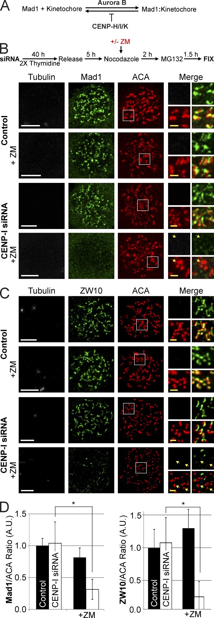
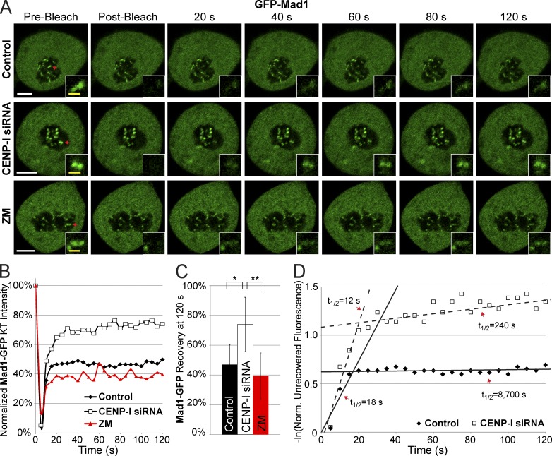
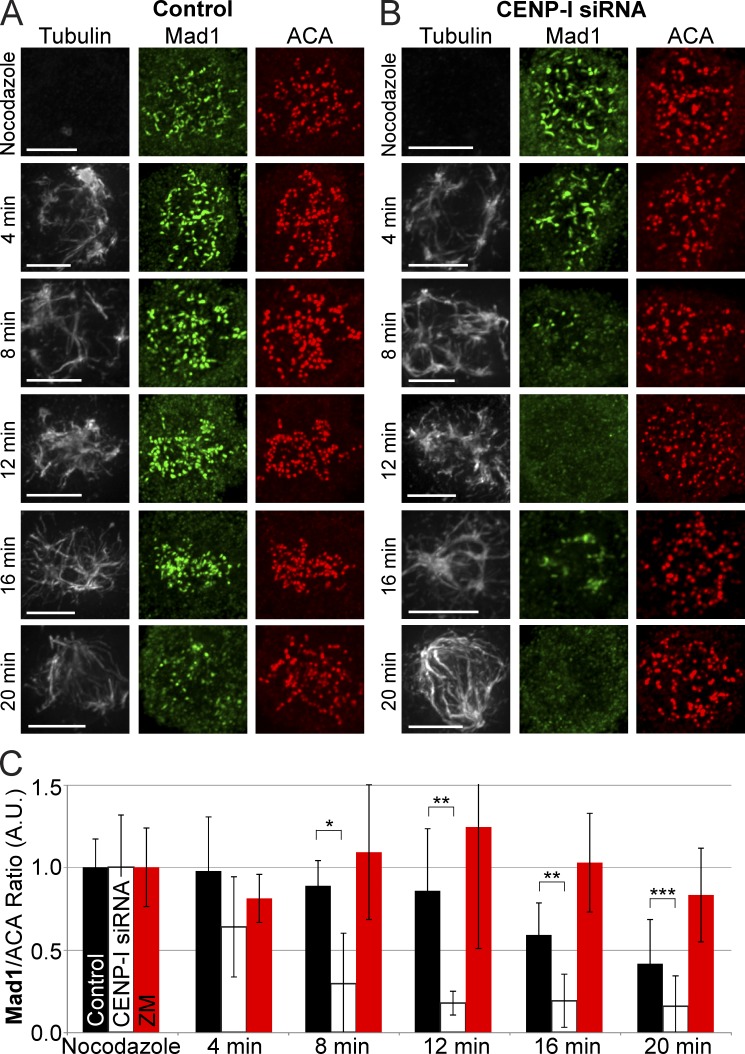
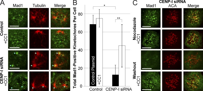
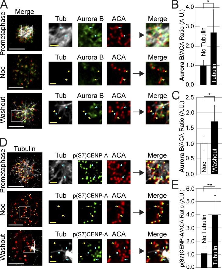
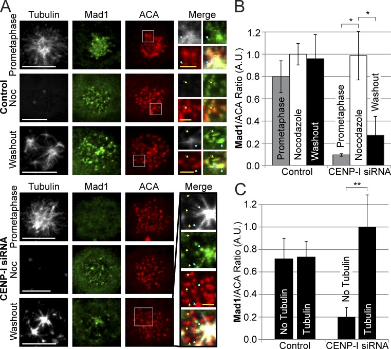
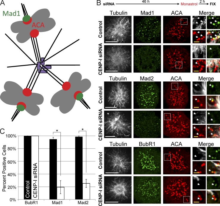
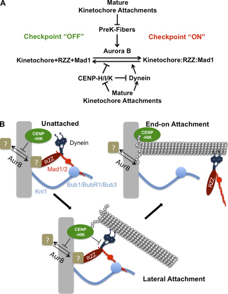
The RZZ (Rod, ZW10, and Zwilch) complex and Mad1 proteins tightly associate with kinetochores to generate the spindle checkpoint signal, but they are released when a kinetochore forms mature microtubule attachments. Here we demonstrate that the centromere protein CENP-I is required to generate a stable association of RZZ and Mad1 with kinetochores. CENP-I also inhibits their removal by dynein stripping. This regulation of Mad1 and RZZ dissociation functions independently of Aurora B, which regulates their association. We show that the microtubule status of each kinetochore independently dictates the recruitment of Aurora B kinase, kinase activity on a kinetochore substrate, and loading of spindle checkpoint proteins. This dynamic regulation of Mad1 association by Aurora B is only uncovered when CENP-I is depleted, consistent with our finding that CENP-I inhibits the dissociation of Mad1. We conclude that the dual activities of Aurora B and CENP-I generate a molecular switch that maintains a robust spindle checkpoint signal at prometaphase kinetochores until they attain mature attachments to microtubules.


Figures

Figure 1.
Figure 1.
Aurora B activity or CENP-I are required to localize Mad1 and ZW10 to unattached kinetochores. (A) Simplified model depicting how Aurora B and CENP-H/I/K function to localize Mad1 at kinetochores. (B and C) Thymidine release assays demonstrating that either CENP-I or Aurora B activity are able to localize Mad1 and ZW10 to unattached kinetochores at the onset of mitosis. After Aurora B inhibition and CENP-I depletion both Mad1 and ZW10 are not at kinetochores. (D) Quantification of Mad1 and ZW10 kinetochore localization from B and C. All cells were treated with 3.3 µM nocodazole. Selected examples of kinetochores without Mad1 or ZW10 are indicated by yellow arrows. Error bars indicate standard deviation. *, P < 0.00005. A.U., arbitrary units. Bars: (white) 5 µm; (yellow) 1 µm.
Figure 2.
Figure 2.
CENP-I increases the half-life of Mad1 at unattached kinetochores. (A) Images of Mad1-GFP FRAP in control, CENP-I–depleted, and ZM-treated cells arrested in nocodazole. (B) Recovery dynamics of Mad1-GFP after photobleaching demonstrating that CENP-I–depleted cells have a larger initial recovery of Mad1 and a faster turnover of stable Mad1. (C) Total recovery of Mad1-GFP at 120 s after photobleaching. (D) Scatter plot displaying the natural log of the normalized unrecovered fluorescence over time. The biphasic nature of Mad1 recovery is illustrated by overlaid lines. CENP-I–depleted cells have a fast phase of initial Mad1 recovery similar to controls but the pool of stable Mad1 in CENP-I–depleted cells has a greatly decreased half-life relative to control. Red arrows in A indicate FRAP targets. FRAP data are from n = 30 experiments. Error bars indicate standard deviation. *, P < 5 × 10⁻⁷; **, P < 2 × 10⁻¹¹. Bars: (white) 5 µm; (yellow) 1 µm.
Figure 3.
Figure 3.
CENP-I–depleted kinetochores lose Mad1 from kinetochores faster than control kinetochores in the presence of microtubules. (A) Control cells retain Mad1 at kinetochores up to 20 min after nocodazole washout. (B) CENP-I–depleted cells lose all Mad1 from kinetochores between 8–12 min after nocodazole washout, even before a bipolar spindle can form. At 16 min, Mad1 can inbriefly be seen at the vertices of microtubule bundles. (C) Quantification of mean Mad1 kinetochore levels across all kinetochores from A and B. Error bars indicate standard deviation. *, P < 10−3; **, P < 10−4; ***, P < 0.05. Indicated statistical significance is between control and CENP-I–depleted groups. A.U., arbitrary units. Bars, 5 µm.
Figure 4.
Figure 4.
CENP-I–depleted kinetochores fail to inhibit dynein-mediated stripping of Mad1. (A) Immunofluorescence images of Mad1 in control and CENP-I–depleted cells 10 min after nocodazole washout, with or without expression of the dynein inhibitor CC1. Control cells retain Mad1 at kinetochores after nocodazole washout, but CENP-I–depleted cells rapidly lose Mad1 from kinetochores and accumulate it at spindle poles in a dynein-dependent manner. (B) Quantification of the total number of Mad1-positive kinetochores in cells from conditions depicted in A. (C) Immunofluorescence images of CENP-I–depleted cells demonstrating that inhibition of dynein does not prevent recruitment of Mad1 to unattached kinetochores, but does prevent loss of Mad1 from kinetochores after nocodazole washout. Centromeres are labeled to demonstrate that Mad1 is at kinetochores. Blue arrows indicate position of spindle poles. Cy5-labeled anti-Mad1 antibody is displayed here in green for ease of viewing. Error bars indicate standard deviation. *, P < 10−7; **, P < 10−3. Bars, 5 µm.
Figure 5.
Figure 5.
Centromere Aurora B localization and activity is enhanced by microtubules at kinetochores. (A) Aurora B localizes to all centromeres during prometaphase and in nocodazole. After nocodazole washout, Aurora B specifically localizes to centromeres where kinetochores overlap with microtubules and is reduced or lost at kinetochores without microtubules. (B) After nocodazole washout Aurora B is specifically enhanced at kinetochores with microtubules and is reduced at kinetochores without microtubules. (C) Quantification of Aurora B centromere intensities in nocodazole and after nocodazole washout demonstrating an increase in overall Aurora B staining across all centromeres after nocodazole washout. (D) Aurora B activity as visualized by p(S7)CENP-A phosphorylation in prometaphase, in nocodazole, and after nocodazole washout. Aurora B activity correlates with the presence of microtubules at kinetochores. (E) p(S7)CENP-A phosphorylation levels are high at kinetochores with microtubules and low at kinetochores without microtubules after nocodazole washout. Yellow arrows indicate select examples of kinetochores without detectable microtubules. Blue arrows indicate select examples of kinetochores with associated microtubules. Each image represents multiple Z-slices. Error bars indicate standard deviation. *, P < 0.05; **, P < 5 × 10⁻⁷. Noc, nocodazole; A.U., arbitrary units. Bars: (white) 5 µm; (yellow) 1 µm.
Figure 6.
Figure 6.
Microtubules trigger Mad1 recruitment to individual kinetochores in CENP-I–depleted cells. (A) Control-depleted cells localize Mad1 to kinetochores in prometaphase, during nocodazole treatment, and 10 min after nocodazole washout. CENP-I–depleted cells have no Mad1 at kinetochores in prometaphase but can fully recruit Mad1 to kinetochores in nocodazole. After nocodazole washout, Mad1 is specifically recruited to kinetochores that overlap with microtubules and is absent from kinetochores without microtubules. (B) Quantification of Mad1 kinetochore intensities from A showing that CENP-I–depleted cells fully recruit Mad1 to unattached kinetochores in nocodazole but lose most Mad1 from kinetochores after nocodazole washout. (C) Quantification of Mad1 intensities at kinetochores with or without microtubules after nocodazole washout. Control cells have equal amounts of Mad1 at kinetochores with or without microtubules, whereas CENP-I–depleted cells have fivefold more Mad1 at kinetochores with microtubules. Yellow arrows indicate select examples of kinetochores without microtubules. Blue arrows indicate select examples of kinetochores with associated microtubules. Insets contain multiple Z-sections for clarity. Error bars indicate standard deviation. *, P < 0.00005; **, P < 0.005. Noc, nocodazole; A.U., arbitrary units. Bars: (white) 5 µm; (yellow) 1 µm.
Figure 7.
Figure 7.
Monastrol-treated cells do not retain Mad1 and Mad2 at anti-poleward kinetochores after CENP-I depletion. (A) Cartoon representation of kinetochore–microtubule attachments in Monastrol-treated cells. (B) Immunofluorescence images of cells after siRNA depletion and Monastrol treatment. Control cells retain Mad1 and Mad2 at anti-poleward kinetochores and recruit BubR1 to all kinetochores. CENP-I–depleted cells can still recruit BubR1 to kinetochores but fail to retain Mad1 and Mad2 at anti-poleward kinetochores. (C) Quantification of B. White arrows indicate select examples of anti-poleward facing kinetochores. Error bars indicate standard deviation. *, P < 0.005. Bars: (white) 5 µm; (yellow) 1 µm.
Figure 8.
Figure 8.
Model for generation and maintenance of the spindle assembly checkpoint. (A) Aurora B activity normally recruits RZZ and Mad1 to unattached kinetochores. PreK-fibers can enhance this process through stimulation of Aurora B activity. CENP-I functions to inhibit their dissociation from kinetochores by stabilizing this interaction and through inhibition of dynein stripping. The formation of mature kinetochore–microtubule attachments suppresses PreK-fibers and extinguishes the stabilizing activity of CENP-I, which allows dynein to strip RZZ and Mad1 from kinetochores. (B) Aurora B and the CENP-H/I/K proteins regulate the recruitment of RZZ by an unknown mechanism, which is shown by recruitment of a receptor that could either be a protein or a posttranslational modification. Kinetochores form early lateral attachments to microtubules through dynein, but the pathway involving CENP-I (CENP-H/I/K) inhibits the dynein-dependent stripping of checkpoint proteins. In this case CENP-I could either inhibit dynein function or make the attachment of RZZ/Mad1 to kinetochores so tight that it can’t be removed by dynein. Upon transition to a mature end-on attachment, CENP-I is turned off and dynein carries checkpoint proteins from kinetochores.

Similar articles

Cited by

References

    1. Amaro A.C., Samora C.P., Holtackers R., Wang E., Kingston I.J., Alonso M., Lampson M., McAinsh A.D., Meraldi P. 2010. Molecular control of kinetochore–microtubule dynamics and chromosome oscillations. Nat. Cell Biol. 12:319–329 10.1038/ncb2033 - DOI - PMC - PubMed
    1. Banerjee B., Kestner C.A., Stukenberg P.T. 2014. EB1 enables spindle microtubules to regulate centromeric recruitment of Aurora B. J. Cell Biol. 204:947–963 10.1083/jcb.201307119 - DOI - PMC - PubMed
    1. Basto R., Gomes R., Karess R.E. 2000. Rough deal and Zw10 are required for the metaphase checkpoint in Drosophila. Nat. Cell Biol. 2:939–943 10.1038/35046592 - DOI - PubMed
    1. Bharadwaj R., Yu H. 2004. The spindle checkpoint, aneuploidy, and cancer. Oncogene. 23:2016–2027 10.1038/sj.onc.1207374 - DOI - PubMed
    1. Burke D.J., Stukenberg P.T. 2008. Linking kinetochore-microtubule binding to the spindle checkpoint. Dev. Cell. 14:474–479 10.1016/j.devcel.2008.03.015 - DOI - PMC - PubMed

Publication types

MeSH terms

LinkOut - more resources