Review
. 2015 Jan;40(1):24-42.
doi: 10.1038/npp.2014.120. Epub 2014 May 27.

Developmental perspectives on oxytocin and vasopressin

Affiliations
Review

Developmental perspectives on oxytocin and vasopressin

Elizabeth A D Hammock. Neuropsychopharmacology. 2015 Jan.

Abstract

The related neuropeptides oxytocin and vasopressin are involved in species-typical behavior, including social recognition behavior, maternal behavior, social bonding, communication, and aggression. A wealth of evidence from animal models demonstrates significant modulation of adult social behavior by both of these neuropeptides and their receptors. Over the last decade, there has been a flood of studies in humans also implicating a role for these neuropeptides in human social behavior. Despite popular assumptions that oxytocin is a molecule of social bonding in the infant brain, less mechanistic research emphasis has been placed on the potential role of these neuropeptides in the developmental emergence of the neural substrates of behavior. This review summarizes what is known and assumed about the developmental influence of these neuropeptides and outlines the important unanswered questions and testable hypotheses. There is tremendous translational need to understand the functions of these neuropeptides in mammalian experience-dependent development of the social brain. The activity of oxytocin and vasopressin during development should inform our understanding of individual, sex, and species differences in social behavior later in life.


Figures

Figure 1
Figure 1
Vasopressin blocks and oxytocin promotes primordial social orienting behaviors in neonatal rodents. Early orienting to social cues increases exposure to social stimuli required for the development of species-specific social behavior expertise. (a) Vasopressin acting at the V1aR eliminates preferences for maternally associated odors in neonatal female mice on postnatal day 8. Copied with permission from Hammock et al (2013). Copyright Elsevier. *Significant difference. (b) Oxytocin antagonism eliminates preferences for maternally associated odors in rats on postnatal day 15. Copied with permission from Kojima and Alberts (2011a). Copyright Elsevier. *Significant huddling preference, Significant difference in huddling preference.
Figure 2
Figure 2
Social contact is associated with oxytocin system activity in rodent and human infants. (a) In neonatal rats, the duration of skin-to-skin contact with a surrogate mother after a period of isolation was positively correlated with the concentration of oxytocin in the hypothalamus. Copied with permission from Kojima et al (2012). Copyright Wiley. (b, c) In 4- to 8-month-old human infants, intranasal oxytocin delivery to their fathers resulted in a very robust increase in infant salivary oxytocin (b) and a significant increase in social gaze and object manipulation in the infants (c), perhaps mediated by enhanced social contact induced by oxytocin in the father (c). Copied with permission from Weisman et al (2012). Copyright Elsevier. *P<0.05, ***P<0.001.
Figure 3
Figure 3
Oxytocin in the neocortex modulates experience-dependent multisensory plasticity. (a) Representative miniature post-synaptic current (mEPSC) recordings for conditions as indicated from the primary somatosensory cortex (S1), primary visual cortex (V1), and primary auditory cortex (Au1). (b) Whisker-deprivation (WD) significantly reduced mEPSC frequencies in S1. (c) WD significantly reduced mEPSC frequencies in V1. (d) WD significantly reduced mEPSC frequencies in Au1. (e) Illustration of the location of the paraventricular (PVN) and supraoptic (SON) nuclei, both in the mouse hypothalamus, in a coronal brain slice. (f) Representative images of oxytocin-positive neurons in the PVN and SON of P14 mice, conditions as indicated; scale bar=200 μm. (g) WD reduced the number of oxytocin-positive neurons in the PVN, but not SON. (h) WD significantly reduced oxytocin peptide level in S1 and V1, but not blood plasma. (i) Oxytocin knockout mice showed reduced mEPSC frequencies in S1. (j) Oxytocin injection increased mEPSC frequencies and rescued the effects of whisker-deprivation in S1. (k) Environmental enrichment (EE) significantly increased oxytocin peptide level in S1 and V1. (l) EE increased mEPSC frequencies and rescued the effects of whisker deprivation in S1. Figure and Figure legend courtesy of Dr Xiang Yu (Zheng et al, 2014), copyright Nature Publishing Group. *P<0.05, **P<0.01, ***P<0.001.
Figure 4
Figure 4
Hypothesis in which development shines light on the ‘Dark Matter of Social Neuroscience’, or the unclarified neural mechanisms between social sensory inputs and social behavior outputs (Insel, 2010). (a) During developmental sensitive periods, parental nurturing (Kojima et al, 2012; Weisman et al, 2012) and/or sensory inputs (Zheng et al, 2014) drive the activity of the PVN resulting in increased release of oxytocin. This oxytocin can regulate multi-sensory plasticity in the neocortex. (b) OXTRs are transiently available during experience-dependent developmental sensitive periods (Hammock and Levitt, 2013). Arrows point to layer II/III of neocortex, which has a high density at postnatal day 14, but not postnatal day 21 or later ages. Perhaps this entrains the developing brain to attend to relevant stimuli, which acquire social definitions because of their proximity in time and space to the nurturing caregiver. After such a developmental trajectory, an adult brain is tuned to ascribe social, affect-modulating properties to multisensory information. (c) For example, wild-type mice are tuned to social stimuli and can rapidly resolve sensory input to determine if a mouse is familiar or novel. In contrast, oxytocin knock-out mice are unable to resolve this social sensory information, despite very active neural processing in primary sensory cortex as measured by c-Fos immunoreactivity after a social encounter (Ferguson et al, 2001), c-Fos images courtesy of Dr Larry Young, copyright The Society for Neuroscience; hatched box at P21 in ‘b’ indicates cortical barrel fields measured in young adult (>P50) mice in panel (c). Thus, developmentally transient experience-dependent neocortical oxytocin signals may allow social support (ie, maternal contact) to fine tune the function of the entire neocortex during a developmental sensitive period for social contact. This layer II/III positioning makes OXTR a promising modulator of multi-sensory integration across modalities by changing the window of opportunity in which separate neural signals could be bound into one unified percept. Developmental modulation in this way by other classic neuroendocrine modulators (eg, CRF, vasopressin) during various sensitive periods may have a significant role in the species-typical development of the brain.

Similar articles

Cited by

References

    1. Aguilar-Valles A, Vaissiere T, Griggs EM, Mikaelsson MA, Takacs IF, Young EJ et al (2013). Methamphetamine-associated memory is regulated by a, writer and an eraser of permissive histone methylation. Biol Psychiatry pii: S0006-3223(13)00855-X. doi:10.1016/j.biopsych.2013.09.014 (e-pub ahead of print). - PMC - PubMed
    1. Albers HE (2012). The regulation of social recognition, social communication and aggression: vasopressin in the social behavior neural network. Horm Behav 61: 283–292. - PubMed
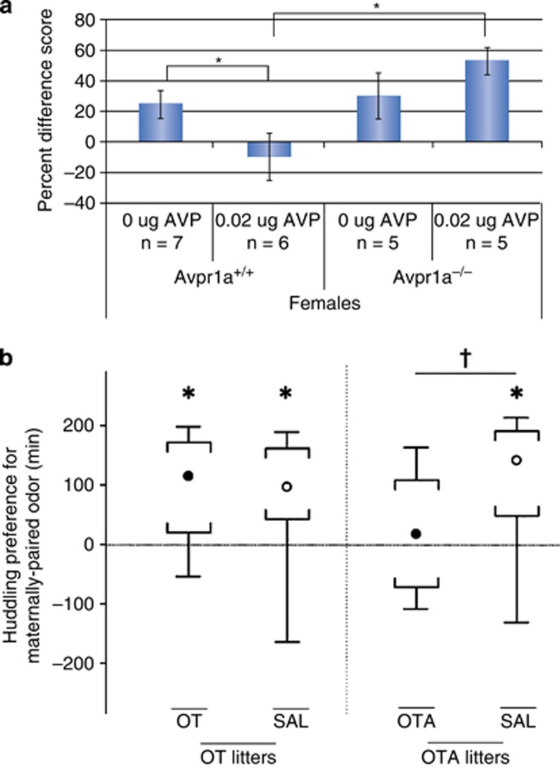
    1. Alberts JR (2007). Huddling by rat pups: ontogeny of individual and group behavior. Dev Psychobiol 49: 22–32 Group huddling behavior responds to central administration of oxytocin on day 10 in neonatal rats. - PubMed
    1. Almazan G, Lefebvre DL, Zingg HH (1989). Ontogeny of hypothalamic vasopressin, oxytocin and somatostatin gene expression. Brain Res Dev Brain Res 45: 69–75. - PubMed
    1. Altstein M, Gainer H (1988). Differential biosynthesis and posttranslational processing of vasopressin and oxytocin in rat brain during embryonic and postnatal development. J Neurosci 8: 3967–3977 This classic paper (among others) demonstrates the earlier emergence of vasopressin relative to oxytocin in rat brain development. - PMC - PubMed

Publication types