. 2014 Jun 2;211(6):1257-70.
doi: 10.1084/jem.20131917. Epub 2014 May 26.

Counter-regulation of T cell effector function by differentially activated p38

Affiliations

Counter-regulation of T cell effector function by differentially activated p38

Muhammad S Alam et al. J Exp Med. .

Abstract

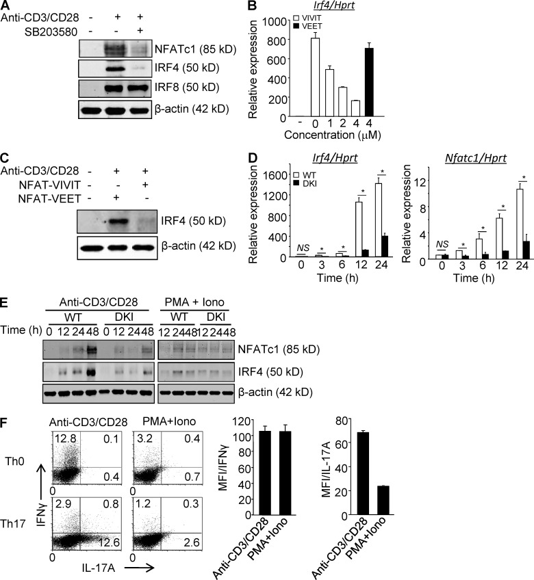
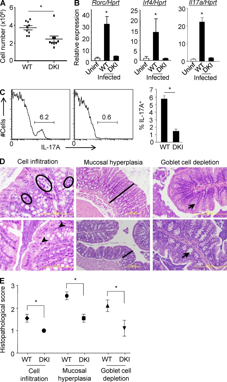
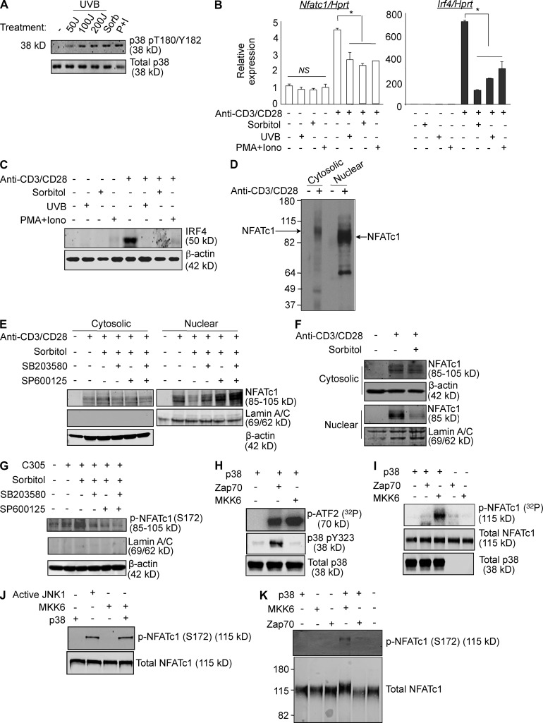
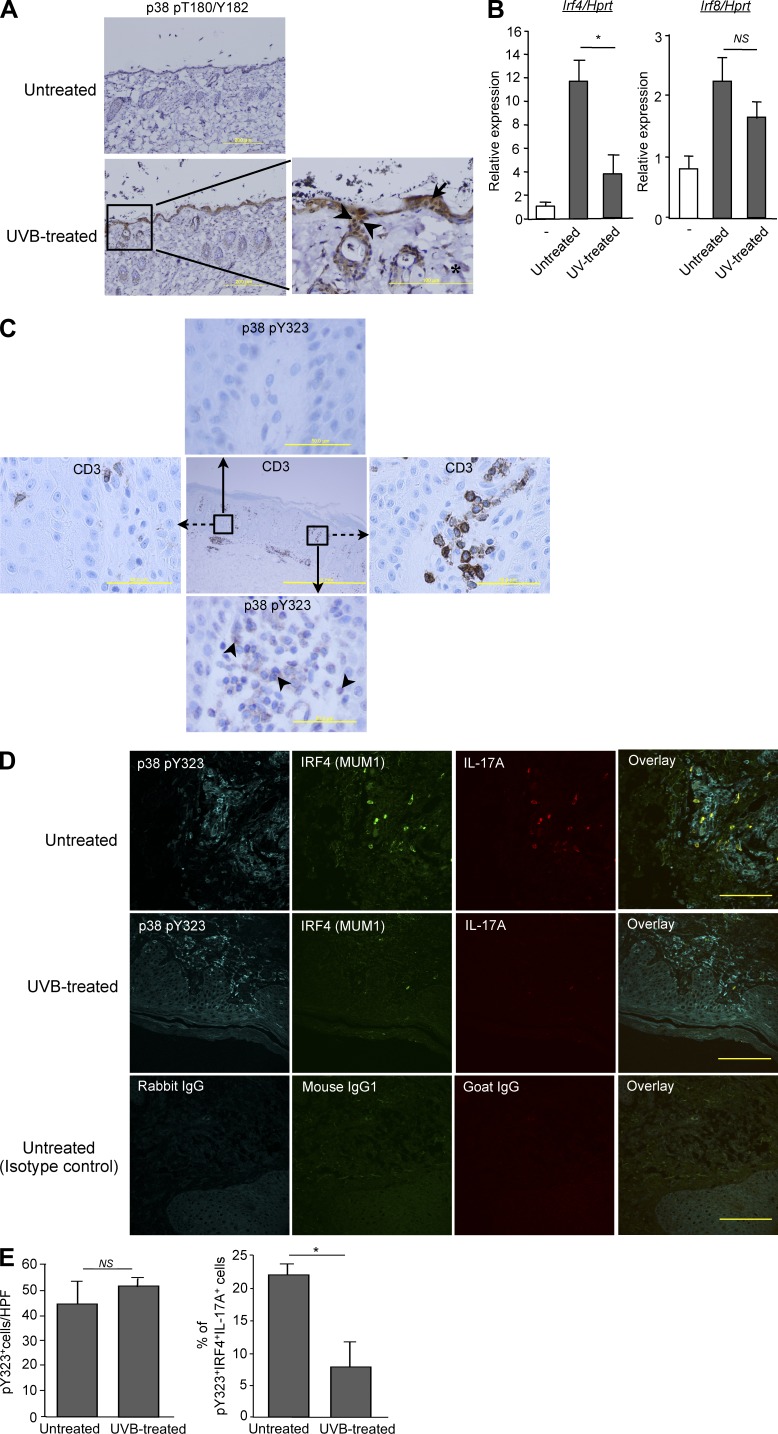
Unlike the MAP kinase (MAPK) cascade that phosphorylates p38 on the activation loop, T cell receptor (TCR) signaling results in phosphorylation on Tyr-323 (pY323, alternative pathway). Using mice expressing p38α and p38β with Y323F substitutions, we show that alternatively but not MAPK cascade-activated p38 up-regulates the transcription factors NFATc1 and IRF4, which are required for proliferation and cytokine production. Conversely, activation of p38 with UV or osmotic shock mitigated TCR-mediated activation by phosphorylation and cytoplasmic retention of NFATc1. Notably, UVB treatment of human psoriatic lesions reduced skin-infiltrating p38 pY323(+) T cell IRF4 and IL-17 production. Thus, distinct mechanisms of p38 activation converge on NFATc1 with opposing effects on T cell immunity, which may underlie the beneficial effect of phototherapy on psoriasis.


Figures

Figure 1.
Figure 1.
p38 Alternative activation is required for TCR-induced expression of NFATc1 and IRF4 in CD4+ T cells. (A) Purified CD4+ T cells were stimulated with anti-CD3/CD28 in the presence or absence of SB203580 and immunoblotted for the indicated transcription factors 48 h later. (B and C) Purified CD4+ T cells were stimulated with anti-CD3/CD28 in the presence of 11R-VIVIT or 11R-VEET. Irf4 mRNA (B) and protein (C) were determined 24 h later. (D and E) Purified CD4+ T cells from WT and DKI mice were stimulated with anti-CD3/CD28 or PMA plus ionomycin, as indicated for the indicated times and analyzed for expression of Nfatc1 and Irf4 mRNA (D) and protein (E). Results shown in A–E are representative of three independent experiments each. (F) Naive CD4+ T cells were stimulated with either anti-CD3/CD28 or PMA and ionomycin under neutral or Th17-skewing conditions for 3 d. IL-17A and IFN-γ expression were determined by intracellular staining and flow cytometry after restimulation with PMA and ionomycin. Results shown in the left panel are representative of three independent experiments and bar graphs in the right panel show the mean ± SEM of all three. *, P < 0.05 (unpaired two-tailed Student’s t test).
Figure 2.
Figure 2.
DKI mice have a defective Th17 response to infection with C. rodentium. (A) WT and DKI mice were infected with C. rodentium by gavage and at day 11–12 cells in the LP were counted. Each symbol represents one mouse, and the mean ± SEM are shown. Data are pooled from two independent experiments with a total of nine mice per group. (B) At day 11 after infection, CD4+ T cells were purified and the expression of Il17a, Rorc, and Irf4 were determined by real-time PCR. The means ± SD are shown (n = 5 mice per group from two independent experiments). (C) IL-17A expression by LP CD4+ T cells was determined by intracellular staining 4 h after stimulation with PMA and ionomycin (left). The mean percentage ± SD of IL-17A–producing CD4+ T cells is shown (right, n = 5 mice per group). Data are representative of two independent experiments. (D) Histological evaluation of representative H&E-stained slides of the colon from WT and DKI mice on day 11 of infection (n = 9 mice per group from 2 independent experiments). Accumulations of lymphocytes are indicated with black circles in WT or arrowheads in DKI mice (left, bar: 100 µm). The thickness of the mucosa is indicated with a black line (middle, bar: 200 µm). Black arrows indicate goblet cells in the colon mucosa (right, bar: 200 µm). (E) The histopathological parameters were quantitated using a double-blinded visual scoring system as detailed in Materials and methods. Data are pooled from 2 independent experiments with a total of 9 mice per group. *, P < 0.05 (unpaired two-tailed Student’s t test).
Figure 3.
Figure 3.
MAPK-cascade activated p38 inhibits functions distal to alternatively activated p38. (A) Purified CD4+ T cells were cultured overnight without stimulation, and then treated with either UV (50 J/m2 or 100 J/m2 or 200 J/m2), 0.6 M sorbitol for 30 min, or PMA (10 ng/ml) and ionomycin (1 µg/ml) for 1 h. Cell lysates were immunoblotted with anti-phospho-p38. The results are representative of three independent experiments. (B) Purified CD4+ T cells were treated with either 0.6 M sorbitol for 30 min, 50 J/m2 UV, or PMA and ionomycin were stimulated with anti-CD3/CD28 for 24 h. The expression of Nfatc1 and Irf4 was determined by quantitative real-time PCR. Results are the mean ± SEM of three independent experiments. *, P < 0.05 (unpaired two-tailed Student’s t test). (C) Cells were treated as in B, and IRF4 expression was determined by Western blot. The results are representative of two independent experiments. (D) Freshly purified T cells were stimulated or not with anti-CD3/CD28 for 48 h. Cytosolic and nuclear fractions were separated in a low percentage SDS-PAGE (8%) gel and immunoblotted for NFATc1. The results are representative of three independent experiments. (E) CD4+ T cells were stimulated for 48 h with anti-CD3/CD28, and then treated with sorbitol in the presence or absence of SB203580 and/or SP600125 for 30 min, rested for 1 h, then immunoblotted for NFATc1 in cytosolic and nuclear fractions. Results are representative of three independent experiments. (F) Purified CD4+ T cells were treated or not with sorbitol for 30 min and stimulated with anti-CD3/CD28 for 48 h, and then immunoblotted for NFATc1 in cytosolic and nuclear fractions. The results are representative of three independent experiments. (G) Jurkat T cells were stimulated for 48 h with an agonistic anti-TCR antibody (C305), and then treated with sorbitol in the presence or absence of SB203580 and/or SP600125 for 30 min. After 1 h without further stimulation, cytosolic fractions were immunoblotted for phospho-NFATc1 (p-NFATc1). The results are representative of three independent experiments. (H) Recombinant p38 was activated with Zap70, MKK6, or buffer alone in in vitro kinase buffer. After 1 h, recombinant ATF2 and 10 µCi [32P]ATP were added for 30 min before separation on SDS-PAGE and PhosphorImager analysis. The phosphorylation state of p38 Y323 was determined by immunoblotting. The results are representative of three independent experiments. (I) Recombinant p38 was activated with Zap70, MKK6, or buffer alone and an in vitro kinase assay using recombinant NFATc1 as substrate was performed as in (H). The results are representative of two independent experiments. (J) Recombinant p38 was activated with MKK6 or buffer alone in in vitro kinase buffer. Active JNK1 was kept in in vitro kinase buffer. After 1 h, recombinant NFATc1 was added and incubated for an additional hour before separation on SDS-PAGE and immunoblotting with an antibody specific for NFATc1 phosphorylated on residue S172. The results are representative of two independent experiments. (K) Recombinant p38 was activated with Zap70, MKK6, or buffer alone in an in vitro kinase assay with recombinant NFATc1 as the p38 substrate, as in I. Samples were separated on a low percentage SDS-PAGE (8%) and immunoblotted for NFATc1 phosphorylated on residue S172. The results are representative of three independent experiments.
Figure 4.
Figure 4.
MAPK cascade–activated p38 inhibits T cell proliferation and effector function mediated by alternatively activated p38. (A and B) Naive CD4+ T cells were treated with 0.6 M sorbitol for 30 min or untreated, and then stimulated with anti-CD3/CD28 in neutral or Th17-skewing conditions. After 2 d, Irf4 and Rorc mRNA levels were analyzed by quantitative real-time PCR (A), and after 3 d, IL-17A expression was determined in cells restimulated with PMA and ionomycin for 4 h. (B) The numbers indicate the frequency of IL-17A–producing cells. The results are representative of two independent experiments. (C and D) Naive CD4+ T cells were treated as in A, and then stimulated with anti-CD3/CD28 either in neutral (Th0) or Th1 skewing conditions for 2 d, at which time Tbet mRNA was measured by real-time PCR (C), and after 3 d cells were restimulated with PMA and ionomycin for 4 h and intracellular IFN-γ expression was measured by flow cytometry (D). The numbers indicate the frequency of IFN-γ–producing cells. *, P < 0.05 (unpaired two-tailed Student’s t test). Data are representative of at least three independent experiments. (E) T cells were stimulated with the indicated concentrations of anti-CD3 in the presence of anti-CD28 (wells coated with 2 µg/ml) or PMA and ionomycin for 48 h and proliferation was assessed by [3H]thymidine incorporation. Data shown are representative of at least three independent experiments.
Figure 5.
Figure 5.
Effect of UV on skin-infiltrating T cells. (A) Mice were exposed or not to UV radiation as described in Materials and methods and, 1 h later, skin sections were obtained and immunostained for CD3 (not depicted) and dual phospho-p38 (pT180/pY182). Phospho-p38–positive T cells are indicated with arrowheads, keratinocytes with arrows, and dermal fibroblasts with asterisks. Bars: 200 µm (left); 100 µm(right). Data are representative of five mice per group from two independent experiments. (B) Purified CD4+ T cells from the skin of UV treated or untreated mice were stimulated in vitro with anti-CD3/CD28 for 24 h, and Irf4 mRNA expression was measured by real-time PCR. Results are the mean ± SEM of two independent experiments with a total of 5 mice per group. *, P < 0.05, NS, not statistically significant (unpaired two-tailed Student’s t test). (C) Immunohistochemical staining of human psoriatic skin lesions with anti-CD3 or phospho-p38 Y323 (p38 pY323)-specific antibodies. The insets in the middle pane show the T cell–poor and T cell–rich areas that are enlarged in the left and right panes for CD3 and the top and bottom panes for p38 pY323. Arrows indicate examples of p38 pY323-positive cells (n = 3 patients; Bars: 1 mm (middle); 50 µm (outer panels). (D) Immunofluorescence staining for p38 pY323 (cyan), IRF4 (green), IL-17A (red), and the corresponding isotype control of representative psoriatic skin lesions biopsies before and after UVB treatment (Bar, 100 µm). (E) Quantitation of p38 pY323+ cells that are also positive for IRF4 and IL-17A (triple positive) in the skin of 6 patients before treatment and 3 of these after UVB treatment. *, P < 0.05. NS, not significant (unpaired two-tailed Student’s t test).

Similar articles

Cited by

References

    1. Aragão A.Z., Nogueira M.L., Granato D.C., Simabuco F.M., Honorato R.V., Hoffman Z., Yokoo S., Laurindo F.R., Squina F.M., Zeri A.C., et al. 2012. Identification of novel interaction between ADAM17 (a disintegrin and metalloprotease 17) and thioredoxin-1. J. Biol. Chem. 287:43071–43082 10.1074/jbc.M112.364513 - DOI - PMC - PubMed
    1. Aujla S.J., Chan Y.R., Zheng M., Fei M., Askew D.J., Pociask D.A., Reinhart T.A., McAllister F., Edeal J., Gaus K., et al. 2008. IL-22 mediates mucosal host defense against Gram-negative bacterial pneumonia. Nat. Med. 14:275–281 10.1038/nm1710 - DOI - PMC - PubMed
    1. Bettelli E., Carrier Y., Gao W., Korn T., Strom T.B., Oukka M., Weiner H.L., Kuchroo V.K. 2006. Reciprocal developmental pathways for the generation of pathogenic effector TH17 and regulatory T cells. Nature. 441:235–238 10.1038/nature04753 - DOI - PubMed
    1. Blomberg K.E., Boucheron N., Lindvall J.M., Yu L., Raberger J., Berglöf A., Ellmeier W., Smith C.E. 2009. Transcriptional signatures of Itk-deficient CD3+, CD4+ and CD8+ T-cells. BMC Genomics. 10:233 10.1186/1471-2164-10-233 - DOI - PMC - PubMed
    1. Brüstle A., Heink S., Huber M., Rosenplänter C., Stadelmann C., Yu P., Arpaia E., Mak T.W., Kamradt T., Lohoff M. 2007. The development of inflammatory T(H)-17 cells requires interferon-regulatory factor 4. Nat. Immunol. 8:958–966 10.1038/ni1500 - DOI - PubMed

Publication types

MeSH terms