

Evolutionarily conserved prefrontal-amygdalar dysfunction in early-life anxiety
- PMID: 24863147
- PMCID: PMC4111803
- DOI: 10.1038/mp.2014.46
Evolutionarily conserved prefrontal-amygdalar dysfunction in early-life anxiety
Abstract
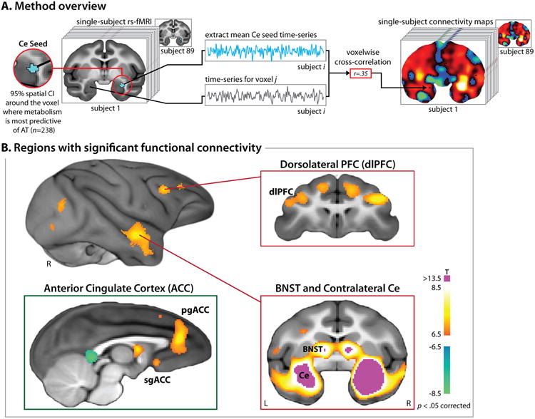
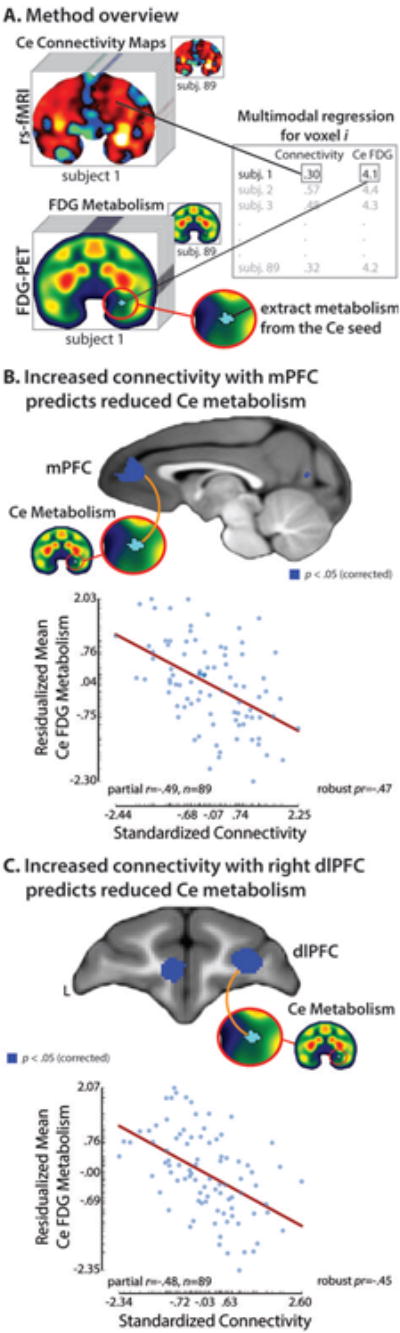
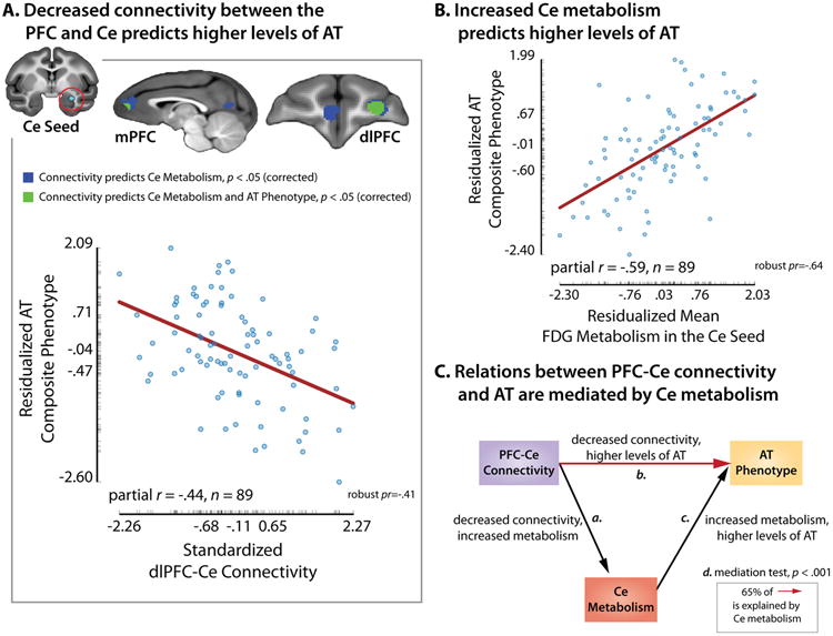
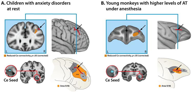
Some individuals are endowed with a biology that renders them more reactive to novelty and potential threat. When extreme, this anxious temperament (AT) confers elevated risk for the development of anxiety, depression and substance abuse. These disorders are highly prevalent, debilitating and can be challenging to treat. The high-risk AT phenotype is expressed similarly in children and young monkeys and mechanistic work demonstrates that the central (Ce) nucleus of the amygdala is an important substrate. Although it is widely believed that the flow of information across the structural network connecting the Ce nucleus to other brain regions underlies primates' capacity for flexibly regulating anxiety, the functional architecture of this network has remained poorly understood. Here we used functional magnetic resonance imaging (fMRI) in anesthetized young monkeys and quietly resting children with anxiety disorders to identify an evolutionarily conserved pattern of functional connectivity relevant to early-life anxiety. Across primate species and levels of awareness, reduced functional connectivity between the dorsolateral prefrontal cortex, a region thought to play a central role in the control of cognition and emotion, and the Ce nucleus was associated with increased anxiety assessed outside the scanner. Importantly, high-resolution 18-fluorodeoxyglucose positron emission tomography imaging provided evidence that elevated Ce nucleus metabolism statistically mediates the association between prefrontal-amygdalar connectivity and elevated anxiety. These results provide new clues about the brain network underlying extreme early-life anxiety and set the stage for mechanistic work aimed at developing improved interventions for pediatric anxiety.
Conflict of interest statement
Authors declare no conflicts of interest.
Figures
References
-
- Darwin C. The expression of the emotions in man and animals. 4th. Oxford University Press; NY: 1872/2009.
-
- Kagan J, Reznick JS, Snidman N. Biological bases of childhood shyness. Science. 1988;240:167–171. - PubMed
-
- Fox NA, Henderson HA, Marshall PJ, Nichols KE, Ghera MM. Behavioral inhibition: linking biology and behavior within a developmental framework. Annu Rev Psychol. 2005;56:235–262. - PubMed
-
- Bystritsky A. Treatment-resistant anxiety disorders. Mol Psychiatry. 2006;11:805–814. - PubMed
Publication types
MeSH terms
Substances
Grants and funding
- MH090912/MH/NIMH NIH HHS/United States
- MH018931/MH/NIMH NIH HHS/United States
- R21 MH091550/MH/NIMH NIH HHS/United States
- P50 MH084051/MH/NIMH NIH HHS/United States
- R21 MH092581/MH/NIMH NIH HHS/United States
- OD011106/OD/NIH HHS/United States
- R01 MH043454/MH/NIMH NIH HHS/United States
- P50 MH100031/MH/NIMH NIH HHS/United States
- MH046729/MH/NIMH NIH HHS/United States
- R01 MH046729/MH/NIMH NIH HHS/United States
- HD008352/HD/NICHD NIH HHS/United States
- RC1 MH090912/MH/NIMH NIH HHS/United States
- P51 RR000167/RR/NCRR NIH HHS/United States
- P30 HD003352/HD/NICHD NIH HHS/United States
- F32 HD008352/HD/NICHD NIH HHS/United States
- MH081884/MH/NIMH NIH HHS/United States
- MH091550/MH/NIMH NIH HHS/United States
- HD003352/HD/NICHD NIH HHS/United States
- R01 MH081884/MH/NIMH NIH HHS/United States
- RR000167/RR/NCRR NIH HHS/United States
- ImNIH/Intramural NIH HHS/United States
- T32 MH018931/MH/NIMH NIH HHS/United States
- MH084051/MH/NIMH NIH HHS/United States
- P51 OD011106/OD/NIH HHS/United States
LinkOut - more resources
Full Text Sources
Other Literature Sources
Medical
