

Transforming growth factor-β-induced lncRNA-Smad7 inhibits apoptosis of mouse breast cancer JygMC(A) cells
- PMID: 24863656
- PMCID: PMC4317863
- DOI: 10.1111/cas.12454
Transforming growth factor-β-induced lncRNA-Smad7 inhibits apoptosis of mouse breast cancer JygMC(A) cells
Abstract
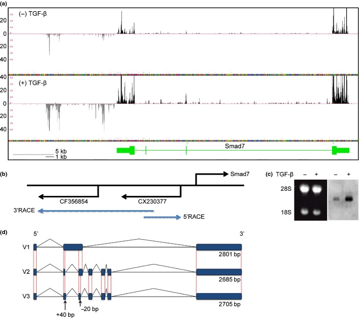
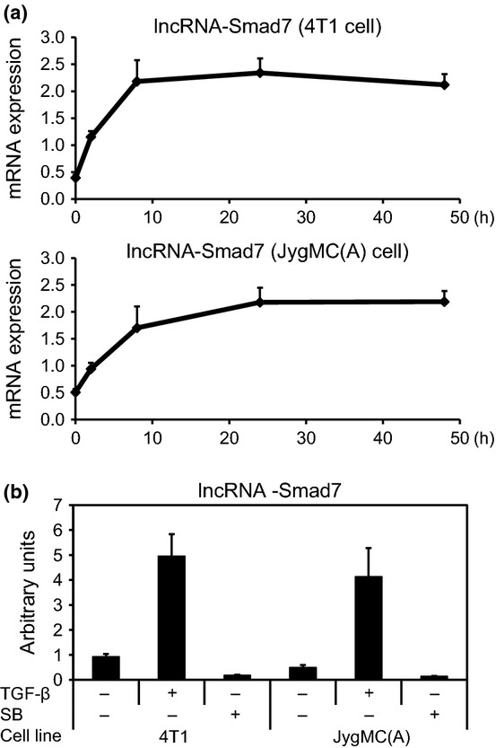
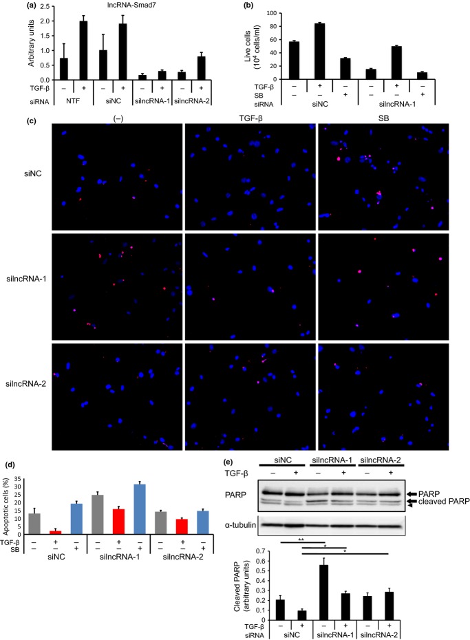
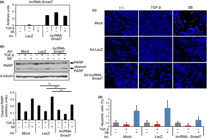
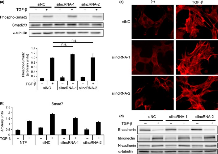
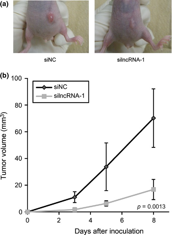
Transforming growth factor (TGF)-β exhibits both pro-apoptotic and anti-apoptotic effects on epithelial cells in a context-dependent manner. The anti-apoptotic function of TGF-β is mediated by several downstream regulatory mechanisms, and has been implicated in the tumor-progressive phenotype of breast cancer cells. We conducted RNA sequencing of mouse mammary gland epithelial (NMuMG) cells and identified a long non-coding RNA, termed lncRNA-Smad7, which has anti-apoptotic functions, as a target of TGF-β. lncRNA-Smad7 was located adjacent to the mouse Smad7 gene, and its expression was induced by TGF-β in all of the mouse mammary gland epithelial cell lines and breast cancer cell lines that we evaluated. Suppression of lncRNA-Smad7 expression cancelled the anti-apoptotic function of TGF-β. In contrast, forced expression of lncRNA-Smad7 rescued apoptosis induced by a TGF-β type I receptor kinase inhibitor in the mouse breast cancer cell line JygMC(A). The anti-apoptotic effect of lncRNA-Smad7 appeared to occur independently of the transcriptional regulation by TGF-β of anti-apoptotic DEC1 and pro-apoptotic Bim proteins. Small interfering RNA for lncRNA-Smad7 did not alter the process of TGF-β-induced epithelial-mesenchymal transition, phosphorylation of Smad2 or expression of the Smad7 gene, suggesting that the contribution of this lncRNA to TGF-β functions may be restricted to apoptosis. Our findings suggest a complex mechanism for regulating the anti-apoptotic and tumor-progressive aspects of TGF-β signaling.
Keywords: Apoptosis; Smad7; breast cancer; long non-coding RNA; transforming growth factor-β.
© 2014 The Authors. Cancer Science published by Wiley Publishing Asia Pty Ltd on behalf of Japanese Cancer Association.
Figures
Similar articles
-
Effect of Smad7 expression on metastasis of mouse mammary carcinoma JygMC(A) cells.J Natl Cancer Inst. 2005 Dec 7;97(23):1734-46. doi: 10.1093/jnci/dji399. J Natl Cancer Inst. 2005. PMID: 16333029
-
Long non-coding RNAs (LncRNA) regulated by transforming growth factor (TGF) β: LncRNA-hit-mediated TGFβ-induced epithelial to mesenchymal transition in mammary epithelia.J Biol Chem. 2015 Mar 13;290(11):6857-67. doi: 10.1074/jbc.M114.610915. Epub 2015 Jan 20. J Biol Chem. 2015. PMID: 25605728 Free PMC article.
-
Activation of extracellular signal-regulated kinase by TGF-beta1 via TbetaRII and Smad7 dependent mechanisms in human bronchial epithelial BEP2D cells.Cell Biol Toxicol. 2007 Mar;23(2):113-28. doi: 10.1007/s10565-006-0097-x. Epub 2006 Nov 9. Cell Biol Toxicol. 2007. PMID: 17096210
-
Current perspectives on inhibitory SMAD7 in health and disease.Crit Rev Biochem Mol Biol. 2020 Dec;55(6):691-715. doi: 10.1080/10409238.2020.1828260. Epub 2020 Oct 20. Crit Rev Biochem Mol Biol. 2020. PMID: 33081543 Review.
-
Expression and function of Smad7 in autoimmune and inflammatory diseases.J Mol Med (Berl). 2021 Sep;99(9):1209-1220. doi: 10.1007/s00109-021-02083-1. Epub 2021 May 31. J Mol Med (Berl). 2021. PMID: 34059951 Free PMC article. Review.
Cited by
-
Mammary Tumor-Associated RNAs Impact Tumor Cell Proliferation, Invasion, and Migration.Cell Rep. 2016 Sep 27;17(1):261-274. doi: 10.1016/j.celrep.2016.08.081. Cell Rep. 2016. PMID: 27681436 Free PMC article.
-
Exosomal lncRNA GAS5 regulates the apoptosis of macrophages and vascular endothelial cells in atherosclerosis.PLoS One. 2017 Sep 25;12(9):e0185406. doi: 10.1371/journal.pone.0185406. eCollection 2017. PLoS One. 2017. PMID: 28945793 Free PMC article.
-
MicroRNA‑96 promotes the proliferation and migration of breast cancer cells by inhibiting Smad7 expression.Oncol Lett. 2025 Jan 22;29(3):151. doi: 10.3892/ol.2025.14897. eCollection 2025 Mar. Oncol Lett. 2025. PMID: 39898288 Free PMC article.
-
Long noncoding RNA NORAD regulates transforming growth factor-β signaling and epithelial-to-mesenchymal transition-like phenotype.Cancer Sci. 2018 Jul;109(7):2211-2220. doi: 10.1111/cas.13626. Epub 2018 May 29. Cancer Sci. 2018. PMID: 29722104 Free PMC article.
-
Long non-coding RNAs and TGF-β signaling in cancer.Cancer Sci. 2020 Aug;111(8):2672-2681. doi: 10.1111/cas.14509. Epub 2020 Jun 17. Cancer Sci. 2020. PMID: 32485023 Free PMC article. Review.
References
-
- Ehata S, Hanyu A, Hayashi M, et al. Transforming growth factor-β promotes survival of mammary carcinoma cells through induction of antiapoptotic transcription factor DEC1. Cancer Res. 2007;67:9694–703. - PubMed
-
- Hoshino Y, Katsuno Y, Ehata S, Miyazono K. Autocrine TGF-β protects breast cancer cells from apoptosis through reduction of BH3-only protein, Bim. J Biochem. 2011;149:55–65. - PubMed
Publication types
MeSH terms
Substances
Associated data
- Actions
- Actions
- Actions
- Actions
LinkOut - more resources
Full Text Sources
Other Literature Sources
Medical
