

Laser light scan analysis of the "anticonvulsant face"
- PMID: 24863698
- PMCID: PMC4246044
- DOI: 10.1002/bdra.23250
Laser light scan analysis of the "anticonvulsant face"
Abstract
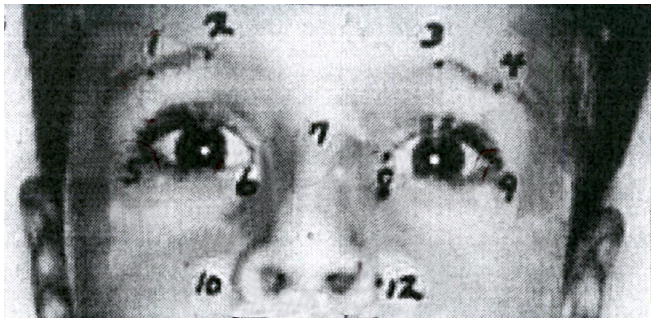
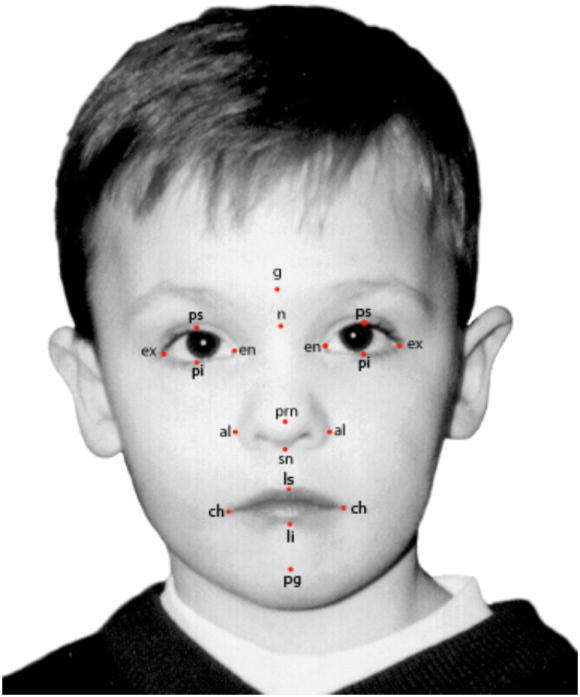
Background: The "anticonvulsant face," with a short nose, broad nasal bridge, epicanthal folds, and wide mouth, was described in the 1970s in children who had been exposed during pregnancy to the anticonvulsant drugs phenytoin and phenobarbital. The laser light scan makes it possible to establish three-dimensional positions of physical features and to determine more objectively the changes in the size and shape of the affected soft tissues of the faces of children exposed to these anticonvulsant drugs during pregnancy.
Methods: Thirteen individuals, exposed throughout pregnancy to phenytoin as either monotherapy or polytherapy, were identified in a previous analysis as having significant changes in their craniofacial features based on measurements of cephalometric radiographs. Those changes were associated with midface hypoplasia and a short nose, features of the "anticonvulsant face." The soft tissues of their faces have been evaluated with laser light scans.
Results: The notable changes in soft tissues identified by laser light scans were a wide philtrum (cph-cph), narrow mouth (ch-ch), short nasal bridge (n-prn), shortened nose height (n-sn), and flattened orbits (orbital protrusion index).
Conclusion: This analysis of the facial features of phenytoin-exposed individuals, selected because of changes in their craniofacial bony structures, showed that there were several significant changes, two of which, widening of the philtrum and a small mouth, have not been described previously as part of this phenotype.
Keywords: anticonvulsants; laser light scanner; structured light scanner; the “anticonvulsant face”.
© 2014 Wiley Periodicals, Inc.
Figures
Similar articles
-
The correlation of deficits in IQ with midface and digit hypoplasia in children exposed in utero to anticonvulsant drugs.J Pediatr. 2005 Jan;146(1):118-22. doi: 10.1016/j.jpeds.2004.08.048. J Pediatr. 2005. PMID: 15644835
-
Facial dysmorphology in children exposed in pregnancy to anticonvulsant medications correlates with deficits in IQ.Am J Med Genet A. 2024 Apr;194(4):e63511. doi: 10.1002/ajmg.a.63511. Epub 2023 Dec 21. Am J Med Genet A. 2024. PMID: 38126162
-
The teratogenicity of anticonvulsant drugs.N Engl J Med. 2001 Apr 12;344(15):1132-8. doi: 10.1056/NEJM200104123441504. N Engl J Med. 2001. PMID: 11297704
-
Prenatal exposure to phenytoin, facial development, and a possible role for vitamin K.Am J Med Genet. 1995 Sep 11;58(3):238-44. doi: 10.1002/ajmg.1320580309. Am J Med Genet. 1995. PMID: 8533825 Review.
-
Drugs in pregnancy: anticonvulsants.Semin Perinatol. 1997 Apr;21(2):114-23. doi: 10.1016/s0146-0005(97)80054-7. Semin Perinatol. 1997. PMID: 9201817 Review.
Cited by
-
Mouth development.Wiley Interdiscip Rev Dev Biol. 2017 Sep;6(5):e275. doi: 10.1002/wdev.275. Epub 2017 May 17. Wiley Interdiscip Rev Dev Biol. 2017. PMID: 28514120 Free PMC article. Review.
-
Correlating Dose and Time of Prenatal Alcohol, Smoking, and Drug Exposure on Craniofacial Morphology: A Systematic Review.Cureus. 2025 Feb 1;17(2):e78320. doi: 10.7759/cureus.78320. eCollection 2025 Feb. Cureus. 2025. PMID: 40034638 Free PMC article. Review.
-
Application of three-dimensional reconstruction technology in dentistry: a narrative review.BMC Oral Health. 2023 Sep 4;23(1):630. doi: 10.1186/s12903-023-03142-4. BMC Oral Health. 2023. PMID: 37667286 Free PMC article. Review.
-
Exposure to Sodium Valproate during Pregnancy: Facial Features and Signs of Autism.Birth Defects Res. 2017 Aug 15;109(14):1134-1143. doi: 10.1002/bdr2.1052. Epub 2017 Jun 21. Birth Defects Res. 2017. PMID: 28635121 Free PMC article.
References
-
- Allanson JE. Objective techniques for craniofacial assessment: what are the choices? Amer J Med Gen. 1997;70:1–5. - PubMed
-
- Altobelli DE. Computer-assisted acquisition of facial surface topography. In: Farkas LG, editor. Anthropometry of head and face. Second edition. Raven Press; New York, NY: 1994. pp. 225–226.
-
- Ardinger HH, Atkin JF, Blackston RD, et al. Verification of the fetal valproate syndrome phenotype. Am J Med Genet. 1988;29:171–185. - PubMed
-
- Astley SJ, Clarren SK. A case definition and photographic screening tool for the facial phenotype of fetal alcohol syndrome. J Pediatr. 1996;129:33–41. - PubMed
-
- Baca DB, Deutsch CK, D’Agostino RB., Jr . Correspondence between direct anthropometry and structured light digit measurement. In: Farkas LG, editor. Anthropometry of the Head and Face. Second edition. Raven Press; New York: 1994. pp. 235–237.
MeSH terms
Substances
Grants and funding
LinkOut - more resources
Full Text Sources
Other Literature Sources
Medical
Research Materials
