Review
. 2014 Jun;143(6):679-92.
doi: 10.1085/jgp.201311153.

Calmodulin regulation (calmodulation) of voltage-gated calcium channels

Affiliations
Review

Calmodulin regulation (calmodulation) of voltage-gated calcium channels

Manu Ben-Johny et al. J Gen Physiol. 2014 Jun.

Abstract

Calmodulin regulation (calmodulation) of the family of voltage-gated CaV1-2 channels comprises a prominent prototype for ion channel regulation, remarkable for its powerful Ca(2+) sensing capabilities, deep in elegant mechanistic lessons, and rich in biological and therapeutic implications. This field thereby resides squarely at the epicenter of Ca(2+) signaling biology, ion channel biophysics, and therapeutic advance. This review summarizes the historical development of ideas in this field, the scope and richly patterned organization of Ca(2+) feedback behaviors encompassed by this system, and the long-standing challenges and recent developments in discerning a molecular basis for calmodulation. We conclude by highlighting the considerable synergy between mechanism, biological insight, and promising therapeutics.


Figures

Figure 1.
Figure 1.
Classic U-shaped signature of CDI. (A–C) Early voltage-clamp recordings from a multicellular preparation of frog atrial trabeculae cells demonstrates CDI of Ca2+ currents. All listed voltages are relative to resting potential ER (∼−80 mV) Adapted from Mentrard et al. (1984). (A) Ca2+ currents (shaded pink) evoked in response to an +80-mV depolarizing test pulse show robust inactivation. Fast capacitive transient was estimated by blocking these Ca2+ currents with 3 mM Co2+ solution (Co). Vertical bar corresponds to 0.5 µA of current; horizontal bar, 100 ms. (B) The Ca2+ current in response to the test pulse is sharply attenuated when preceded by an +80 mV prepulse. This reduction in current amplitude reflects the inactivation of Ca2+ channels. Format is as in A. (C) Further increase in the prepulse potential to +120 mV restores Ca2+ current amplitude (compare with B). Here, the diminished Ca2+ entry during the prepulse was insufficient to trigger CDI. The format is as in A. (D–F) Single channel recordings of L-type Ca2+ channels from adult rat ventricular myocytes also exhibit U-shaped dependence of Ca2+ channel inactivation. Adapted from Imredy and Yue (1994) with permission from Elsevier. (D) Depolarizing voltage pulse to +20 mV evokes representative elementary Ca2+ currents. Ensemble average currents are shown on the bottom. (E) When depolarizing pulse was preceded by +40 mV prepulse, the elementary Ca2+ currents are sparser and the first opening is delayed. This reduction in open probability parallels the sharp attenuation of macroscopic Ca2+ currents seen in B. The ensemble average is shown on the bottom. (F) Further increase in prepulse voltage to +120 mV led to a reversal of this gating pattern, once again highlighting the U-shaped dependence of CDI. Ensemble average is shown on the bottom.
Figure 2.
Figure 2.
Calmodulation: the ingredients and the flavors. (A) Channel diagram illustrates overall arrangement of structural landmarks critical for CDI. The Ca2+-inactivation (CI) region, spanning ∼160 residues of the channel carboxy terminus, is highly conserved across CaV1/2 channel families and is elemental for CDI. The proximal segment (PCI) of the CI region includes the dual vestigial EF hand (EF) segments (shaded purple and blue-green). The IQ domain, a canonical CaM binding motif critical for CDI, is located just downstream of the PCI segment. The NSCaTE element in the amino terminus of CaV1.2/1.3 channels is an N-lobe Ca2+/CaM effector site. The CBD element (gray) in the carboxy terminus of CaV2 channels is a CaM binding segment thought to be critical for CDI. (B) The table outlines functional bipartition of CaM in CaV channels and the corresponding spatial Ca2+ selectivities. In CaV1.2 and CaV1.3 channels, both the C-lobe and N-lobe of CaM enable fast CDI with local Ca2+ selectivity. Latent CDI of CaV1.4 channels is revealed upon deletion of an autoinhibitory domain in the distal carboxy terminus. Throughout the CaV2 channel family, the N-lobe of CaM evokes slow CDI with global Ca2+ spatial selectivity. In CaV2.1 channels, the C-lobe of CaM supports ultra-fast facilitation (CDF) with local Ca2+ selectivity. No C-lobe triggered modulation has been reported for CaV2.2 and CaV2.3 channels.
Figure 3.
Figure 3.
Toward an atomic-level understanding of CDI. (A) IQ-centric view of calmodulation of CaV channels. In this mechanistic scheme, ApoCaM is preassociated with the IQ domain (blue). Upon Ca2+ binding, CaM rebinds the same IQ domain with a higher affinity, and subtle conformational rearrangements are presumed to trigger CaM-mediated channel regulation. (B, left) Crystal structure of CaV1.2 IQ domain peptide in complex with Ca2+/CaM (Protein Data Bank accession no. 2BE6). The IQ domain is colored blue. CaM is show in cyan (N-lobe, pale cyan; C-lobe, cyan). Ca2+ ions are depicted as yellow spheres. The key isoleucine residue (red) serves as a hydrophobic anchor for CaM. Ca2+/CaM adopts a parallel arrangement with the IQ domain in which the N-lobe binds closer to the amino terminus of the IQ domain and the C-lobe binds further downstream. (B, right) Crystal structure of Ca2+/CaM bound to mutant CaV1.2 IQ domain with alanines substituted for the key isoleucine-glutamine residues (accession no. 2F3Z). This double mutation abolishes CDI. Structurally, however, Ca2+/CaM hugs the mutant IQ domain in a similar conformation as to its interaction with the wild-type (left). (C) Crystal structure of Ca2+/CaM bound to CaV2.1 IQ domain (left, accession no. 3BXK; right, accession no. 3DVM). The IQ domain is shaded green, CaM in cyan. Ca2+/CaM was reported to bind to the CaV2.1 IQ domain in both parallel (left) and antiparallel (right) arrangements. The antiparallel arrangement in which the C-lobe of CaM binds upstream of IQ domain has led to speculation that CDF may result from this inverted polarity of Ca2+/CaM association. (D) Simplified configurations of the CaM/channel complex relevant for calmodulation (E, A, IC, IN, and ICN). In configuration E, channels lack preassociated CaM and therefore cannot undergo CDI. Configuration A corresponds to channels bound to apoCaM, and thus capable of undergoing robust CDI. Ca2+ binding to the C-lobe and N-lobe of CaM leads to the inactivated configurations IC and IN, respectively. Ca2+ binding to both lobes of CaM then yields configuration ICN, with both C and N lobes of CaM engaged toward CDI. This final transition may exhibit positive cooperativity as specified by the constant λ >> 1. Under endogenous levels of CaM, channels may reside in any of the five configurations. Strong coexpression of wild-type or mutant CaM restricts accessibility to various states. Reproduced with permission from Ben Johny et al., 2013.
Figure 4.
Figure 4.
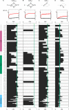
Residue-level roadmap for calmodulation of CaV1.3 channels. Systematic alanine scanning mutagenesis of the entire CI domain of CaV1.3 channels. (A) CDI of wild-type and mutant CaV1.3 channels recombinantly expressed in HEK293 cells was measured with only endogenous CaM present. CDI observed here reflects properties of the entire system (stick figure) diagrammed in Fig. 3 D. Exemplar whole-cell current shows robust CDI for wild-type (WT) CaV1.3, reflecting their high affinity for apoCaM. Here and throughout, the vertical bar pertains to 0.2 nA of Ca2+ current (black); the Ba2+ current (gray) has been scaled ∼3-fold downward to aid comparison of decay kinetics. Horizontal bar, 100 ms. CDI is measured as the fractional reduction of Ca2+ current in comparison to Ba2+ at 300 ms (Ben Johny et al., 2013). Bottom, bar graph summary of CDI for alanine substitutions for indicated residues. CDI for WT channels, blue-green vertical line. Alanine substitutions in both EF hand regions and IQ domain resulted in diminished CDI. (B) Overexpression of CaMWT isolates the behavior of the diamond-shaped subsystem. Such overexpression of CaMWT should rescue CDI for mutations that weakened apoCaM preassociation. For WT CaV1.3, exemplar current shows that CDI is unaltered, as expected for a channel with high affinity for apoCaM. Bottom, bar graph summary of CDI for various mutant channels with CaMWT overexpressed. Format is as in A. CDI is rescued for several mutations in the EF hand region (EF2) and the IQ domain, reflecting the preassociation of N-lobe and C-lobe of apoCaM. (C) Overexpression of CaM12 isolates C-lobe CDI. Exemplar currents for WT channels show the fast C-lobe form of CDI. Bottom, bar graph summary shows the footprint of C-lobe CDI. Mutants in both the first EF hand and the IQ domain disrupted the C-lobe CDI (see the LGF loci in the EF hand regions and TFL and IQD loci in the IQ domain). The format is as in A. (D) Overexpression of CaM34 isolates N-lobe CDI. N-lobe CDI was largely preserved by mutations in the CI region. Mutations that weakened N-lobe apoCaM preassociation exhibited enhanced N-lobe CDI (see EF2 segment). Reassuringly, alanine scanning mutagenesis of NSCaTE element resulted in specific disruption of N-lobe CDI of CaV1.3 channels (Tadross et al., 2008). Adapted with permission from Ben Johny et al. (2013) and Bazzazi et al. (2013).
Figure 5.
Figure 5.
Next-generation blueprint for CaM/CaBP regulation of CaV channels. (A) De novo molecular model of CaV1.3 CI region docked to apoCaM. The N-lobe of apoCaM is thought to preassociate with the PCI region (green), whereas the C-lobe binds to the IQ domain (blue). The NSCaTE segment (tan) is unoccupied. This model corresponds to configuration A in Fig. 3 D, where the channels are charged with an apoCaM and ready to undergo CDI. (B) Proposed model for the Ca2+ inactivated state of the channel. The N-lobe of Ca2+/CaM is shown binding to the NSCaTE segment based on a recent NMR structure (Protein Data Bank accession no. 2LQC). This configuration is believed to result in N-lobe CDI. The de novo molecular model shows C-lobe of Ca2+/CaM, the EF hand segment, and the IQ domain forming a tripartite complex resulting in C-lobe CDI. Overall, this model corresponds to the inactive configuration ICN in Fig. 3 D. (C) Proposed allosteric mechanism of CaBP action. CaBP and apoCaM may dually bind the CaV1 channel. However, CaBP may allosterically inhibit CDI of CaV1 channels by interacting with the NT, III-IV loop, or PCI segment, thereby preventing Ca2+/CaM from reaching its effector configuration. (A–C) Adapted with permission from Yang et al. (2014).

References

    1. Adams P.J., Rungta R.L., Garcia E., van den Maagdenberg A.M., MacVicar B.A., Snutch T.P. 2010. Contribution of calcium-dependent facilitation to synaptic plasticity revealed by migraine mutations in the P/Q-type calcium channel. Proc. Natl. Acad. Sci. USA. 107:18694–18699 10.1073/pnas.1009500107 - DOI - PMC - PubMed
    1. Alseikhan B.A., DeMaria C.D., Colecraft H.M., Yue D.T. 2002. Engineered calmodulins reveal the unexpected eminence of Ca2+ channel inactivation in controlling heart excitation. Proc. Natl. Acad. Sci. USA. 99:17185–17190 10.1073/pnas.262372999 - DOI - PMC - PubMed
    1. Anderson M.E., Mohler P.J. 2009. Rescuing a failing heart: think globally, treat locally. Nat. Med. 15:25–26 10.1038/nm0109-25 - DOI - PubMed
    1. Ashcroft F.M., Stanfield P.R. 1981. Calcium dependence of the inactivation of calcium currents in skeletal muscle fibers of an insect. Science. 213:224–226 10.1126/science.213.4504.224 - DOI - PubMed
    1. Babitch J. 1990. Channel hands. Nature. 346:321–322 10.1038/346321b0 - DOI - PubMed

Publication types