. 2014 Jun;143(6):783-97.
doi: 10.1085/jgp.201311125.

Impact of detubulation on force and kinetics of cardiac muscle contraction

Affiliations

Impact of detubulation on force and kinetics of cardiac muscle contraction

Cecilia Ferrantini et al. J Gen Physiol. 2014 Jun.

Abstract

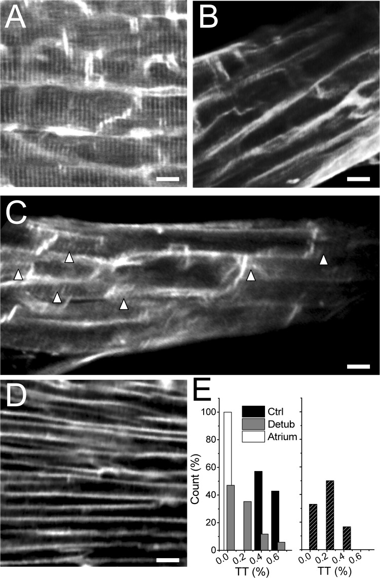
Action potential-driven Ca(2+) currents from the transverse tubules (t-tubules) trigger synchronous Ca(2+) release from the sarcoplasmic reticulum of cardiomyocytes. Loss of t-tubules has been reported in cardiac diseases, including heart failure, but the effect of uncoupling t-tubules from the sarcolemma on cardiac muscle mechanics remains largely unknown. We dissected intact rat right ventricular trabeculae and compared force, sarcomere length, and intracellular Ca(2+) in control trabeculae with trabeculae in which the t-tubules were uncoupled from the plasma membrane by formamide-induced osmotic shock (detubulation). We verified disconnection of a consistent fraction of t-tubules from the sarcolemma by two-photon fluorescence imaging of FM4-64-labeled membranes and by the absence of tubular action potential, which was recorded by random access multiphoton microscopy in combination with a voltage-sensitive dye (Di-4-AN(F)EPPTEA). Detubulation reduced the amplitude and prolonged the duration of Ca(2+) transients, leading to slower kinetics of force generation and relaxation and reduced twitch tension (1 Hz, 30°C, 1.5 mM [Ca(2+)]o). No mechanical changes were observed in rat left atrial trabeculae after formamide shock, consistent with the lack of t-tubules in rodent atrial myocytes. Detubulation diminished the rate-dependent increase of Ca(2+)-transient amplitude and twitch force. However, maximal twitch tension at high [Ca(2+)]o or in post-rest potentiated beats was unaffected, although contraction kinetics were slower. The ryanodine receptor (RyR)2 Ca-sensitizing agent caffeine (200 µM), which increases the velocity of transverse Ca(2+) release propagation in detubulated cardiomyocytes, rescued the depressed contractile force and the slower twitch kinetics of detubulated trabeculae, with negligible effects in controls. We conclude that partial loss of t-tubules leads to myocardial contractile abnormalities that can be rescued by enhancing and accelerating the propagation of Ca(2+)-induced Ca(2+) release to orphan RyR2 clusters.


Figures

Figure 1.
Figure 1.
Transient volume, SL, and force changes during formamide-induced osmotic shock. (A) Representative stereomicroscope image of a rat right ventricular trabecula mounted between a force transducer (left, basket) and a motor (right, hook). The highlighted region is displayed below as observed with a 20× microscope objective to show trabecular diameter changes occurring during formamide exposure. Bars, 50 µm. (B; top) Changes in muscle volume versus time during formamide osmotic shock protocol. 1.5 M formamide was applied for 20 min and rapidly removed. Muscle volume is estimated from trabecular width, thickness, and length by assuming the trabeculae to be elliptical cylinders. Values are means ± SE from six trabeculae. (Bottom) SL versus time during formamide shock; means ± SE from seven rat ventricular trabeculae. (C) Representative twitch force changes during the experimental protocol. Twitch active stress increases to a maximum within the first 3 min of formamide exposure (on average by 146 ± 63% in six trabeculae). Twitch force is above the basal level during the entire formamide exposure period and shows a further transient increase during the first 30 s of formamide washout. Subsequently, twitch force decreases to a minimum (47 ± 24% of preexposure level) before attaining a new steady level in 7–8 min. Several mechanisms may contribute to the transient force increase during formamide exposure (e.g., [Ca2+]i increase and reduced myofilament spacing caused by cell shrinkage, direct effects of formamide on force generation, etc.). However, it appears to be a rather transitory and unspecific event because it occurs both in formamide-treated ventricular and atrial trabeculae (Fig. 5).
Figure 2.
Figure 2.
Formamide shock is able to effectively induce acute detubulation of thin rat ventricular trabeculae. (A) TPF image of a control formamide-untreated ventricular trabecula stained with FM4-64, showing a uniform pattern of t-tubules. (B and C) TPF image of ventricular trabeculae stained with FM4-64 after the detubulation protocol. The loss of TT after formamide shock is incomplete: Δ in C indicates residual TT after formamide shock. (D) TPF image of a FM4-64–stained atrial trabecula, in which most of the myocytes show the absence of TT. Bars in A–D, 10 µm. (E; left) Frequency distribution of mean TT component (TT%) obtained by averaging TT% values from six different areas in each trabecula. (Right) Frequency distribution of TT% values in six different areas of a representative detubulated ventricular trabecula. TT% was quantified by FFT analysis of the image. Although the mean TT% is statistically reduced in formamide-treated trabeculae, the effects of formamide shock between different areas can be variable within a single trabecula, ranging from a complete absence of connected T-tubules (TT% of ∼0) to minimal TT disconnection (TT% of >0.4).
Figure 3.
Figure 3.
Optical recordings of surface sarcolemma and t-tubule transmembrane voltage confirm TT detachment after formamide shock. (A) TPF image of a Di-4-AN(F)EPPTEA–stained rat ventricular trabecula. Transmembrane voltage at multiple membrane sites was measured with a RAMP microscope. The lines mark the probed sarcolemma regions: surface sarcolemma (SS) is red, and t-tubule (TT) is green. (B) Normalized fluorescence traces from the scanned lines indicated in A. TT and surface sarcolemma action potentials are identical. (C) TPF image of a rat ventricular trabecula after formamide shock: the trabecula was stained before the detubulation procedure. (D) Normalized fluorescence traces from the scanned lines indicated in C: reduced florescence signal is detected in the scanned TATS region, suggesting an electrical uncoupling from surface sarcolemma. (E) Mean ± SE data of fluorescence signal amplitude and action potential duration (in msec) at 50% (APD50%) and 90% (APD90%) of repolarization. Bars, 10 µm. **, P < 0.01, paired.
Figure 4.
Figure 4.
Mechanical consequences of acute detubulation in ventricular trabeculae: prolongation of contraction kinetics and impaired positive force–frequency response. (A) Representative traces of isometric twitch force and corresponding [Ca2+]i transient at 1- and 3-Hz stimulation frequency before (Control) and after (Detubulation) formamide osmotic shock. [Ca2+]i was monitored during contraction after loading the trabeculae with fura-2 AM. (B) Steady-state force–frequency relationship and frequency dependency of twitch kinetics in control and detubulated ventricular trabeculae. Data points in the plots represent mean ± SE data of active stress (left), twitch peak time (middle), and 90% relaxation time (right) at frequencies from 0.1 to 8 Hz. Extracellular [Ca2+] was equal to 1.5 mM. n = 8 and 12, number of rats and number of trabeculae, respectively. #, P < 0.01, paired t test for frequencies from 1 to 8 Hz; **, P < 0.01, paired t test at all frequencies; *, P < 0.05, paired t test at all frequencies.
Figure 5.
Figure 5.
Formamide osmotic shock does not affect mechanical properties of atrial myocardium. (A) TPF images of a rat atrial trabecula loaded with FM4-64. The lack of organized t-tubules is evident. Bars, 10 µm. (B) Transient changes in twitch tension during formamide exposure and washout. Twitch tension shows a significant increase in the presence of formamide, a further enhancement during the initial phase of formamide washout, followed by a rapid decrease below basal levels. All of these changes are comparable to those observed in ventricular trabeculae during formamide treatment. However, in atrial trabeculae, twitch tension recovers basal pre-formamide amplitude and kinetics some minutes after formamide shock. (C) Superimposed representative traces of isometric twitch force at 1-Hz stimulation frequency from an atrial trabecula before and after formamide shock. Extracellular [Ca2+]i is equal to 1.5 mM.
Figure 6.
Figure 6.
Effects of acute detubulation on maximal twitch force in post-rest potentiated beats and high extracellular [Ca2+]. (A) Representative traces of isometric twitch force after 30-s stimulation pauses in a trabecula before and after formamide treatment. (Inset) Superimposed Ca2+ transients at baseline (0.5 Hz; dotted lines) and after pauses (continuous lines) before and after detubulation (black and gray, respectively). (B) Mean ± SE data of twitch tension and kinetics after 30-s pauses (Post Rest) and at 8 mM of extracellular [Ca2+] (High [Ca]). **, P < 0.01, paired. n = 10 and 13, number of rats and number of trabeculae, respectively.
Figure 7.
Figure 7.
Low-dose caffeine selectively improves twitch force and contractile kinetics in detubulated trabeculae. (A) Representative traces showing steady-state twitches at 3 Hz (left) and corresponding [Ca2+]i transients (right) in a rat trabecula before and after formamide shock, at baseline (gray and black) and in the presence of 200 µmol/L caffeine (blue and red). (Inset) Force–frequency relationship in the same muscle showing that the effect of caffeine on twitch tension after detubulation was more prominent at a high frequency. (B) Twitch and [Ca2+]i amplitude at baseline and in the presence of 200 µmol/L caffeine. (C) Twitch kinetics and [Ca2+]i transient kinetics at baseline and in the presence of 200 µmol/L caffeine. Means ± SE from seven rat ventricular trabeculae (four rats). *, P < 0.05; **, P < 0.01, paired, caffeine versus baseline.
Figure 8.
Figure 8.
Low-dose caffeine improves CICR transverse propagation velocity in detubulated myocytes. (A) Representative confocal line scan images of Fluoforte-loaded cardiomyocytes showing steady-state Ca2+ transients at 1 Hz, 1 mM [Ca2+]o, and room temperature. (B) Mean data of CICR transverse propagation velocity in detubulated myocytes in the absence and presence of 200 µM caffeine. Velocity was taken from the slope of the Ca2+ wave in line scan images (dotted lines in yellow). Data are mean ± SE. The XY scale bar is 100 ms × 10 µm. **, P < 0.01, unpaired. The total number of myocytes tested was: 40 in the control group, 71 in the detubulated group, and 54 in the detubulated plus caffeine group. Myocytes were isolated from four rat hearts.

References

    1. Allegra Mascaro A.L., Sacconi L., Pavone F.S. 2010. Multi-photon nanosurgery in live brain. Front Neuroenergetics. 2:21. - PMC - PubMed
    1. Bassani R.A., Bers D.M. 1994. Na-Ca exchange is required for rest-decay but not for rest-potentiation of twitches in rabbit and rat ventricular myocytes. J. Mol. Cell. Cardiol. 26:1335–1347 10.1006/jmcc.1994.1152 - DOI - PubMed
    1. Baudino T.A., Carver W., Giles W., Borg T.K. 2006. Cardiac fibroblasts: friend or foe? Am. J. Physiol. Heart Circ. Physiol. 291:H1015–H1026 10.1152/ajpheart.00023.2006 - DOI - PubMed
    1. Bers D.M., Bassani R.A., Bassani J.W., Baudet S., Hryshko L.V. 1993. Paradoxical twitch potentiation after rest in cardiac muscle: Increased fractional release of SR calcium. J. Mol. Cell. Cardiol. 25:1047–1057 10.1006/jmcc.1993.1117 - DOI - PubMed
    1. Brette F., Komukai K., Orchard C.H. 2002. Validation of formamide as a detubulation agent in isolated rat cardiac cells. Am. J. Physiol. Heart Circ. Physiol. 283:H1720–H1728 - PubMed

Publication types