

Distribution of protein poly(ADP-ribosyl)ation systems across all domains of life
- PMID: 24865146
- PMCID: PMC4245714
- DOI: 10.1016/j.dnarep.2014.05.003
Distribution of protein poly(ADP-ribosyl)ation systems across all domains of life
Abstract
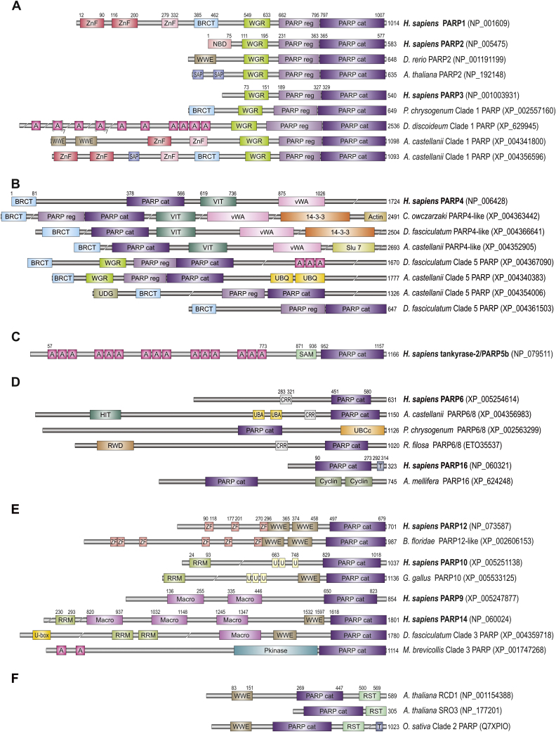
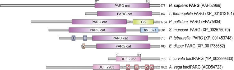
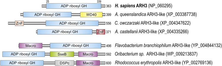
Poly(ADP-ribosyl)ation is a post-translational modification of proteins involved in regulation of many cellular pathways. Poly(ADP-ribose) (PAR) consists of chains of repeating ADP-ribose nucleotide units and is synthesized by the family of enzymes called poly(ADP-ribose) polymerases (PARPs). This modification can be removed by the hydrolytic action of poly(ADP-ribose) glycohydrolase (PARG) and ADP-ribosylhydrolase 3 (ARH3). Hydrolytic activity of macrodomain proteins (MacroD1, MacroD2 and TARG1) is responsible for the removal of terminal ADP-ribose unit and for complete reversion of protein ADP-ribosylation. Poly(ADP-ribosyl)ation is widely utilized in eukaryotes and PARPs are present in representatives from all six major eukaryotic supergroups, with only a small number of eukaryotic species that do not possess PARP genes. The last common ancestor of all eukaryotes possessed at least five types of PARP proteins that include both mono and poly(ADP-ribosyl) transferases. Distribution of PARGs strictly follows the distribution of PARP proteins in eukaryotic species. At least one of the macrodomain proteins that hydrolyse terminal ADP-ribose is also always present. Therefore, we can presume that the last common ancestor of all eukaryotes possessed a fully functional and reversible PAR metabolism and that PAR signalling provided the conditions essential for survival of the ancestral eukaryote in its ancient environment. PARP proteins are far less prevalent in bacteria and were probably gained through horizontal gene transfer. Only eleven bacterial species possess all proteins essential for a functional PAR metabolism, although it is not known whether PAR metabolism is truly functional in bacteria. Several dsDNA viruses also possess PARP homologues, while no PARP proteins have been identified in any archaeal genome. Our analysis of the distribution of enzymes involved in PAR metabolism provides insight into the evolution of these important signalling systems, as well as providing the basis for selection of the appropriate genetic model organisms to study the physiology of the specific human PARP proteins.
Keywords: DNA damage response; Macrodomain; PARG; PARP; Poly(ADP-ribose).
Copyright © 2014 The Authors. Published by Elsevier B.V. All rights reserved.
Figures
References
-
- Gibson B.A., Kraus W.L. New insights into the molecular and cellular functions of poly(ADP-ribose) and PARPs. Nat. Rev. Mol. Cell Biol. 2012;13:411–424. - PubMed
-
- Barkauskaite E., Jankevicius G., Ladurner A.G., Ahel I., Timinszky G. The recognition and removal of cellular poly(ADP-ribose) signals. FEBS J. 2013;280:3491–3507. - PubMed
-
- Hottiger M.O., Hassa P.O., Luscher B., Schuler H., Koch-Nolte F. Toward a unified nomenclature for mammalian ADP-ribosyltransferases. Trends Biochem. Sci. 2010;35:208–219. - PubMed
-
- Tao Z., Gao P., Liu H.W. Identification of the ADP-ribosylation sites in the PARP-1 automodification domain: analysis and implications. J. Am. Chem. Soc. 2009;131:14258–14260. - PubMed
Publication types
MeSH terms
Substances
Grants and funding
LinkOut - more resources
Full Text Sources
Other Literature Sources
