

Myocardial CT perfusion imaging and SPECT for the diagnosis of coronary artery disease: a head-to-head comparison from the CORE320 multicenter diagnostic performance study
- PMID: 24865312
- PMCID: PMC4263655
- DOI: 10.1148/radiol.14140806
Myocardial CT perfusion imaging and SPECT for the diagnosis of coronary artery disease: a head-to-head comparison from the CORE320 multicenter diagnostic performance study
Erratum in
-
Myocardial CT Perfusion Imaging and SPECT for the Diagnosis of Coronary Artery Disease: A Head-to-Head Comparison from the CORE320 Multicenter Diagnostic Performance Study.Radiology. 2015 Feb;274(2):626. doi: 10.1148/radiol.14144050. Radiology. 2015. PMID: 25625749 Free PMC article. No abstract available.
Abstract
Purpose: To compare the diagnostic performance of myocardial computed tomographic (CT) perfusion imaging and single photon emission computed tomography (SPECT) perfusion imaging in the diagnosis of anatomically significant coronary artery disease (CAD) as depicted at invasive coronary angiography.
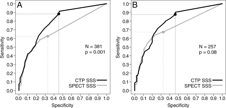
Materials and methods: This study was approved by the institutional review board. Written informed consent was obtained from all patients. Sixteen centers enrolled 381 patients from November 2009 to July 2011. Patients underwent rest and adenosine stress CT perfusion imaging and rest and either exercise or pharmacologic stress SPECT before and within 60 days of coronary angiography. Images from CT perfusion imaging, SPECT, and coronary angiography were interpreted at blinded, independent core laboratories. The primary diagnostic parameter was the area under the receiver operating characteristic curve (Az). Sensitivity and specificity were calculated with use of prespecified cutoffs. The reference standard was a stenosis of at least 50% at coronary angiography as determined with quantitative methods.
Results: CAD was diagnosed in 229 of the 381 patients (60%). The per-patient sensitivity and specificity for the diagnosis of CAD (stenosis ≥50%) were 88% (202 of 229 patients) and 55% (83 of 152 patients), respectively, for CT perfusion imaging and 62% (143 of 229 patients) and 67% (102 of 152 patients) for SPECT, with Az values of 0.78 (95% confidence interval: 0.74, 0.82) and 0.69 (95% confidence interval: 0.64, 0.74) (P = .001). The sensitivity of CT perfusion imaging for single- and multivessel CAD was higher than that of SPECT, with sensitivities for left main, three-vessel, two-vessel, and one-vessel disease of 92%, 92%, 89%, and 83%, respectively, for CT perfusion imaging and 75%, 79%, 68%, and 41%, respectively, for SPECT.
Conclusion: The overall performance of myocardial CT perfusion imaging in the diagnosis of anatomic CAD (stenosis ≥50%), as demonstrated with the Az, was higher than that of SPECT and was driven in part by the higher sensitivity for left main and multivessel disease.
Figures
Comment in
-
Comparing Apples and Oranges: High-End Cardiac 320-Detector Row CT vs Archaic SPECT Protocols.Radiology. 2015 May;275(2):620-2. doi: 10.1148/radiol.2015142007. Radiology. 2015. PMID: 25906309 No abstract available.
Similar articles
-
Prognostic Value of Combined CT Angiography and Myocardial Perfusion Imaging versus Invasive Coronary Angiography and Nuclear Stress Perfusion Imaging in the Prediction of Major Adverse Cardiovascular Events: The CORE320 Multicenter Study.Radiology. 2017 Jul;284(1):55-65. doi: 10.1148/radiol.2017161565. Epub 2017 Mar 14. Radiology. 2017. PMID: 28290782 Free PMC article.
-
Accuracy of Computed Tomographic Angiography and Single-Photon Emission Computed Tomography-Acquired Myocardial Perfusion Imaging for the Diagnosis of Coronary Artery Disease.Circ Cardiovasc Imaging. 2015 Oct;8(10):e003533. doi: 10.1161/CIRCIMAGING.115.003533. Circ Cardiovasc Imaging. 2015. PMID: 26467105 Free PMC article. Clinical Trial.
-
Diagnostic accuracy of combined coronary angiography and adenosine stress myocardial perfusion imaging using 320-detector computed tomography: pilot study.Eur Radiol. 2013 Jul;23(7):1812-21. doi: 10.1007/s00330-013-2788-z. Epub 2013 Feb 21. Eur Radiol. 2013. PMID: 23430194
-
Diagnostic Performance of Hybrid Cardiac Imaging Methods for Assessment of Obstructive Coronary Artery Disease Compared With Stand-Alone Coronary Computed Tomography Angiography: A Meta-Analysis.JACC Cardiovasc Imaging. 2018 Apr;11(4):589-599. doi: 10.1016/j.jcmg.2017.05.020. Epub 2017 Aug 16. JACC Cardiovasc Imaging. 2018. PMID: 28823745 Free PMC article.
-
Advances in Single-Photon Emission Computed Tomography: Hardware, Software, and Myocardial Flow Reserve.Heart Fail Clin. 2025 Jul;21(3):327-337. doi: 10.1016/j.hfc.2025.01.018. Heart Fail Clin. 2025. PMID: 40506147 Review.
Cited by
-
Stress echocardiography: what is new and how does it compare with myocardial perfusion imaging and other modalities?Curr Cardiol Rep. 2015 Jun;17(6):43. doi: 10.1007/s11886-015-0600-1. Curr Cardiol Rep. 2015. PMID: 25911442 Review.
-
Clinical Applications of Wide-Detector CT Scanners for Cardiothoracic Imaging: An Update.Korean J Radiol. 2019 Dec;20(12):1583-1596. doi: 10.3348/kjr.2019.0327. Korean J Radiol. 2019. PMID: 31854147 Free PMC article. Review.
-
Clinical applications of cardiac computed tomography: a consensus paper of the European Association of Cardiovascular Imaging-part II.Eur Heart J Cardiovasc Imaging. 2022 Mar 22;23(4):e136-e161. doi: 10.1093/ehjci/jeab292. Eur Heart J Cardiovasc Imaging. 2022. PMID: 35175348 Free PMC article.
-
CT myocardial perfusion imaging: current status and future perspectives.Int J Cardiovasc Imaging. 2017 Jul;33(7):1009-1020. doi: 10.1007/s10554-017-1102-6. Epub 2017 Mar 9. Int J Cardiovasc Imaging. 2017. PMID: 28281025 Review.
-
Evidence for myocardial CT perfusion imaging in the diagnosis of hemodynamically significant coronary artery disease.Cardiovasc Diagn Ther. 2015 Feb;5(1):58-62. doi: 10.3978/j.issn.2223-3652.2015.01.03. Cardiovasc Diagn Ther. 2015. PMID: 25774349 Free PMC article.
References
-
- George RT, Arbab-Zadeh A, Miller JM, et al. . Adenosine stress 64- and 256-row detector computed tomography angiography and perfusion imaging: a pilot study evaluating the transmural extent of perfusion abnormalities to predict atherosclerosis causing myocardial ischemia. Circ Cardiovasc Imaging 2009;2(3):174–182. - PMC - PubMed
-
- George RT, Arbab-Zadeh A, Miller JM, et al. . Computed tomography myocardial perfusion imaging with 320-row detector computed tomography accurately detects myocardial ischemia in patients with obstructive coronary artery disease. Circ Cardiovasc Imaging 2012;5(3):333–340. - PubMed
-
- Blankstein R, Shturman LD, Rogers IS, et al. . Adenosine-induced stress myocardial perfusion imaging using dual-source cardiac computed tomography. J Am Coll Cardiol 2009;54(12):1072–1084. - PubMed
-
- Bettencourt N, Chiribiri A, Schuster A, et al. . Direct comparison of cardiac magnetic resonance and multidetector computed tomography stress-rest perfusion imaging for detection of coronary artery disease. J Am Coll Cardiol 2013;61(10):1099–1107. - PubMed
Publication types
MeSH terms
Grants and funding
LinkOut - more resources
Full Text Sources
Other Literature Sources
Medical
Miscellaneous
