. 2014 Jul;20(7):1078-89.
doi: 10.1261/rna.045187.114. Epub 2014 May 27.

Biochemical defects in minor spliceosome function in the developmental disorder MOPD I

Affiliations

Biochemical defects in minor spliceosome function in the developmental disorder MOPD I

Faegheh Jafarifar et al. RNA. 2014 Jul.

Abstract

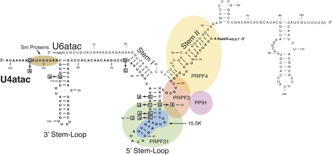
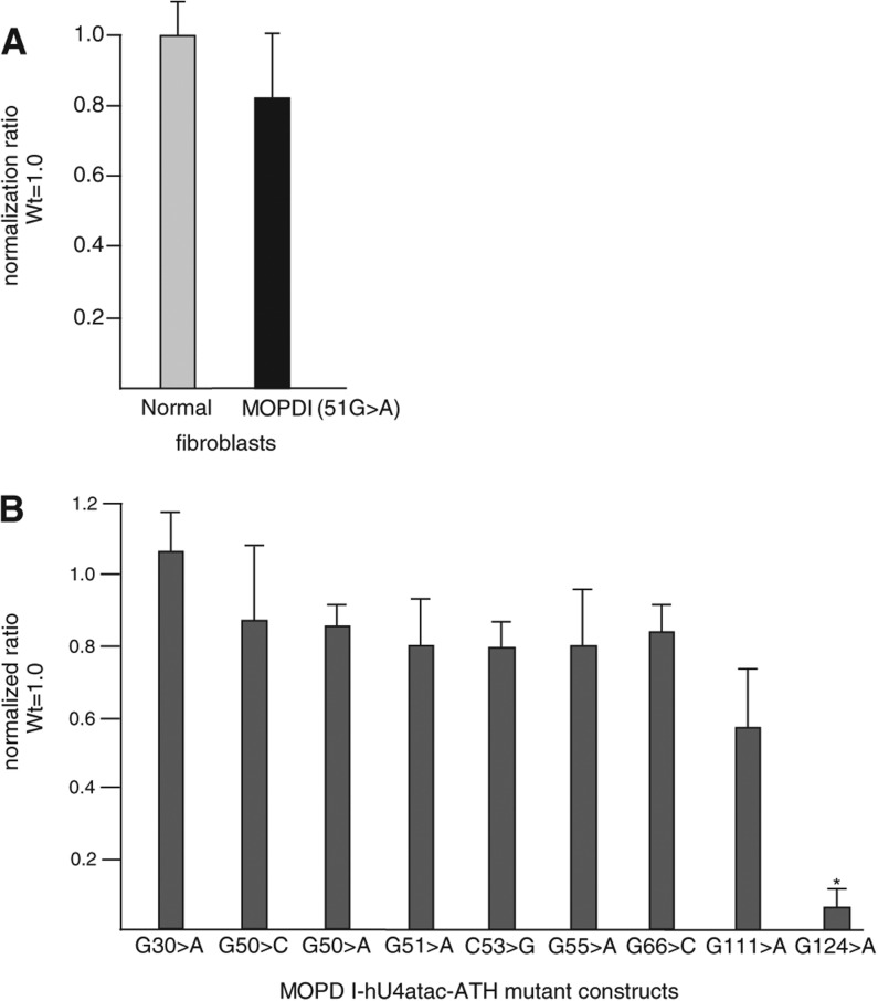
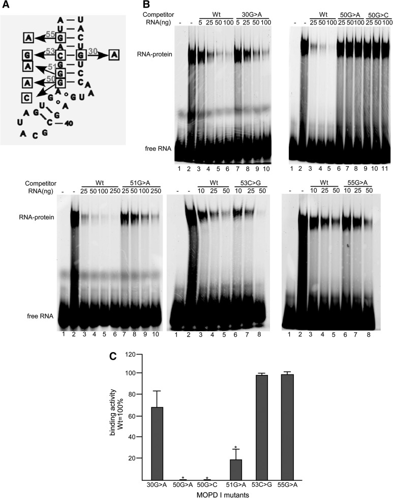
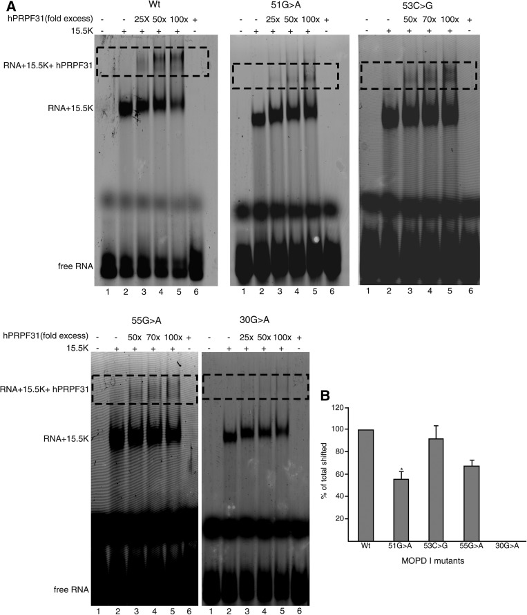
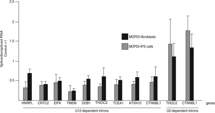
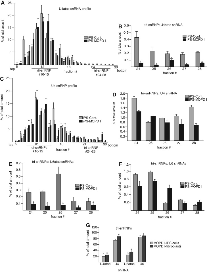
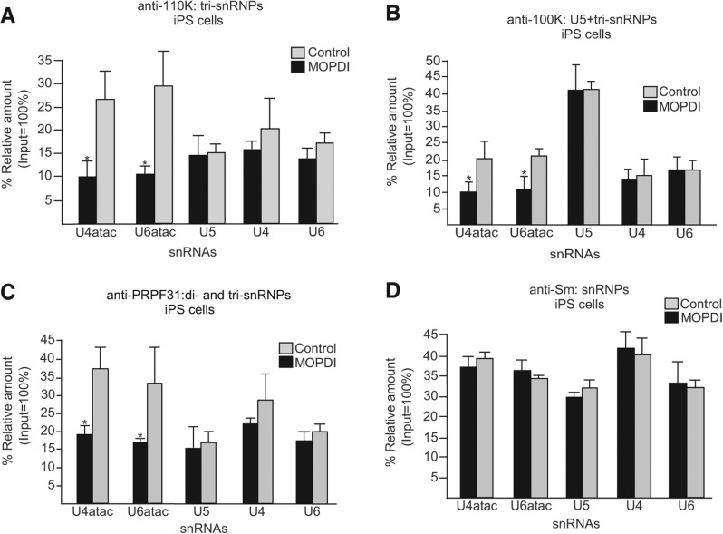
Biallelic mutations of the human RNU4ATAC gene, which codes for the minor spliceosomal U4atac snRNA, cause the developmental disorder, MOPD I/TALS. To date, nine separate mutations in RNU4ATAC have been identified in MOPD I patients. Evidence suggests that all of these mutations lead to abrogation of U4atac snRNA function and impaired minor intron splicing. However, the molecular basis of these effects is unknown. Here, we use a variety of in vitro and in vivo assays to address this question. We find that only one mutation, 124G>A, leads to significantly reduced expression of U4atac snRNA, whereas four mutations, 30G>A, 50G>A, 50G>C and 51G>A, show impaired binding of essential protein components of the U4atac/U6atac di-snRNP in vitro and in vivo. Analysis of MOPD I patient fibroblasts and iPS cells homozygous for the most common mutation, 51G>A, shows reduced levels of the U4atac/U6atac.U5 tri-snRNP complex as determined by glycerol gradient sedimentation and immunoprecipitation. In this report, we establish a mechanistic basis for MOPD I disease and show that the inefficient splicing of genes containing U12-dependent introns in patient cells is due to defects in minor tri-snRNP formation, and the MOPD I-associated RNU4ATAC mutations can affect multiple facets of minor snRNA function.

Keywords: U4atac snRNA; disease; snRNP function; splicing.


Figures

FIGURE 1.
FIGURE 1.
Diagram of the human U4atac/U6atac di-snRNP. U4atac snRNA (black) is shown base-paired with U6atac snRNA (gray), forming a Y-shaped functional structure that interacts with several essential spliceosomal proteins. The intermolecular stem II domain interacts with three proteins: PRPF4 (yellow), PPIH (purple), and PRPF3 (orange). The U4atac intramolecular 5′ stem–loop domain interacts with the 15.5K (blue) and PRPF31 (green) proteins. PRPF31 directly interacts with PRPF3. A specific sequence located at the 3′ end of U4atac snRNA binds the Sm protein complex (brown). The nucleotides that are mutated in MOPD I patients are boxed.
FIGURE 2.
FIGURE 2.
Steady-state levels of mutant U4atac snRNAs. (A) The amounts of U4atac snRNA in normal and homozygous 51G>A MOPD I umbilical cord fibroblast cells were determined by real-time quantitative RT-PCR after isolation of total RNAs. U6 snRNA was used for normalization. The amount of U4atac snRNA in normal cells was set to unity. (B) The amounts of MOPD I-associated mutant U4atac snRNA were determined by real-time quantitative RT-PCR after isolation of total RNAs from CHO cells cotransfected with plasmids expressing either modified wild-type human U4atac snRNA or MOPD I mutant U4atac snRNA as well as modified U6atac snRNA, which is able to base-pair with the modified U4atac snRNA. Primers were designed to amplify only the transfected snRNAs. The level of cotransfected human U12 snRNA was used for normalization. The amount of transfected wild-type human U4atac snRNA was set to unity. Results are expressed as mean and one standard deviation from three independent experiments. (*) A significant difference, P < 0.05, using a two-tailed t-test.
FIGURE 3.
FIGURE 3.
Binding activity of the 15.5K protein to the 5′ stem–loop of MOPD I U4atac snRNAs. (A) Positions of MOPD I-associated mutations in the intramolecular 5′ stem–loop of human U4atac snRNA. (B) Electrophoretic gel shift analysis of 15.5K protein binding to labeled wild-type RNA in the presence of various amounts of unlabeled competitor RNA. In each panel, lane 1 is labeled wild-type RNA only and lane 2 is labeled wild-type RNA with 15.5K protein. Additional lanes contain the amounts of the various unlabeled competitor RNAs as listed: (Wt) wild type. (C) Quantitation of competition activity compared to wild-type RNA, which was set to 100%. Results are expressed as mean and one standard deviation for three experiments. (*) Indicates a significant difference, P < 0.05, using a two-tailed t-test.
FIGURE 4.
FIGURE 4.
Binding of the PRPF31 protein to 5′ stem–loop RNA-15.5K protein complexes. (A) Labeled wild type (Wt) or mutant U4atac 5′ stem–loop RNAs were incubated with 15.5K protein and different amounts of PRPF31 protein. In each panel, lane 1 is RNA only; lane 2 is RNA plus 15.5K protein; lane 6 is RNA plus PRPF31 protein, and lanes 35 are reactions containing the listed fold excess of PRPF31 protein to the RNA-15.5K complex. The positions of free RNA, RNA-15.5K complex, and PRPF31-15.5K-RNA complex (dotted regions), are indicated in the figure. (B) Plot showing the percentage amounts of supershifted RNA-15.5K complex. Using ImageJ, the amounts of RNA shifted by 15.5K protein and the amounts of RNA supershifted (dotted region) by 15.5K + PRPF31 proteins were measured for each titration. The percentage of supershifted RNAs was calculated for each titration and normalized to wild type, which was set to 100%.
FIGURE 5.
FIGURE 5.
Analysis of 51G>A MOPD I-derived iPS cells. Endogenous U12-dependent introns are inefficiently spliced in iPS cells to similar extents as in MOPD I fibroblast cells. The ratio of spliced to unspliced pre-mRNA for U12- and U2-dependent introns was determined by real-time quantitative RT-PCR as described (He et al. 2011). Two MOPD I-derived iPS cell lines were compared to two MOPD I fibroblast cell lines. For each intron, the ratio seen in control fibroblasts or control iPS cells is set to 1.0. Although U12-dependent introns show increased retention in both MOPD I-iPS and MOPD I fibroblast cell lines, U2-dependent introns are unaffected in both cell lines.
FIGURE 6.
FIGURE 6.
Glycerol gradient sedimentation and characterization of snRNP profiles in 51G>A MOPD I fibroblasts and iPS cell lines. (A) Overall distribution of U4atac snRNA in control and MOPD I iPS cell lines. Whole-cell extracts from control and 51G>A MOPD I-derived iPS cell lines were applied to a 10%–30% glycerol gradient, and snRNP species were separated in different fractions. Total RNA was extracted from each fraction, and U4atac snRNA levels were examined with quantitative RT-PCR by using specific primers for U4atac snRNA. The abundance of U4atac snRNA in each fraction (percentage of total) was calculated as the amount of U4atac snRNA in each fraction divided by the sum of the amount in all fractions. The fraction numbers and the positions of di- and tri-snRNP complexes are shown at the bottom. The direction of sedimentation is from top (left) to bottom (right). (B) Detail of the tri-snRNP fractions shown in A. Results are expressed as mean and one standard deviation from three independent experiments. (C) The overall distribution of U4 snRNA in control and MOPD I iPS cell lines was measured in the same fractions as in A. (D) Detail of U4 snRNA in tri-snRNP fractions. (E) Detail of U6atac snRNA in tri-snRNP fractions. (F) Detail of U6 snRNA in tri-snRNP fractions. (G) An identical analysis was performed using extracts from control and patient fibroblast cells, and the amounts of various snRNAs in the tri-snRNP fractions were compared with the results using control and MOPD I iPS cells. The amounts of the various snRNAs in control cell tri-snRNPs were set to 100%.
FIGURE 7.
FIGURE 7.
Immunoprecipitation analysis of snRNPs. (A) Antibody against the 110K tri-snRNP-specific protein was used to immunoprecipitate snRNPs from extracts prepared from control and MOPD I-derived iPS cells. The amounts of immunoprecipitated snRNAs were then measured by quantitative RT-PCR using specific primers for each minor and major snRNA. The amount of input RNA was set to 100%. (B) Immunoprecipitation using antibodies against the 100K U5-specific protein, which is also a component of tri-snRNPs. (C) Immunoprecipitation using antibodies against the PRPF31 di- and tri-snRNP-specific protein. (D) Immunoprecipitation using antibodies against the Sm proteins. Results are expressed as mean and one standard deviation from three independent experiments. (*) Indicates a significant difference, P < 0.05, two-tailed t-test.

References

    1. Abdel-Salam GMH, Miyake N, Eid MM, Abdel-Hamid MS, Hassan NA, Eid OM, Effat LK, El-Badry TH, El-Kamah GY, El-Darouti M, et al. 2011. A homozygous mutation in RNU4ATAC as a cause of microcephalic osteodysplastic primordial dwarfism type I (MOPD I) with associated pigmentary disorder. Am J Med Genet A 155A: 2885–2896 - PubMed
    1. Abdel-Salam GM, Abdel-Hamid MS, Issa M, Magdy A, El-Kotoury A, Amr K 2012. Expanding the phenotypic and mutational spectrum in microcephalic osteodysplastic primordial dwarfism type I. Am J Med Genet A 158A: 1455–1461 - PubMed
    1. Abdel-Salam GM, Abdel-Hamid MS, Hassan NA, Issa MY, Effat L, Ismail S, Aglan MS, Zaki MS 2013. Further delineation of the clinical spectrum in RNU4ATAC related microcephalic osteodysplastic primordial dwarfism type I. Am J Med Genet A 161A: 1875–1881 - PubMed
    1. Alioto TS 2007. U12DB: A database of orthologous U12-type spliceosomal introns. Nucleic Acids Res 35: D110–D115 - PMC - PubMed
    1. Boulisfane N, Choleza M, Rage F, Neel H, Soret J, Bordonné R 2011. Impaired minor tri-snRNP assembly generates differential splicing defects of U12-type introns in lymphoblasts derived from a type I SMA patient. Hum Mol Genet 20: 641–648 - PubMed

Publication types

MeSH terms

Supplementary concepts

LinkOut - more resources