. 2014 Aug;34(15):2874-89.
doi: 10.1128/MCB.00135-14. Epub 2014 May 27.

PZR coordinates Shp2 Noonan and LEOPARD syndrome signaling in zebrafish and mice

Affiliations

PZR coordinates Shp2 Noonan and LEOPARD syndrome signaling in zebrafish and mice

Jeroen Paardekooper Overman et al. Mol Cell Biol. 2014 Aug.

Abstract

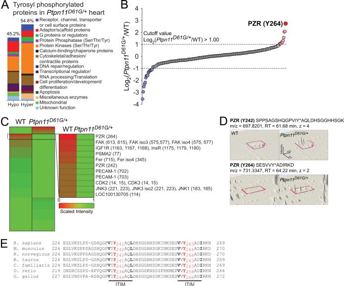
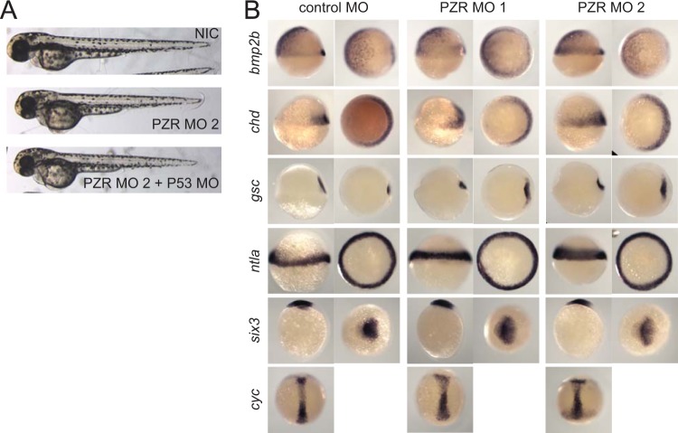
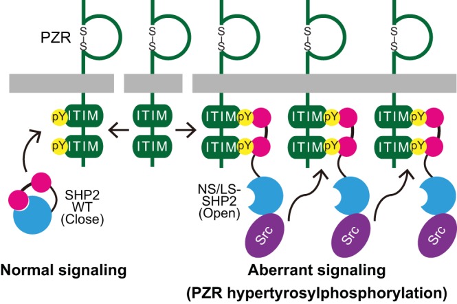
Noonan syndrome (NS) is an autosomal dominant disorder caused by activating mutations in the PTPN11 gene encoding Shp2, which manifests in congenital heart disease, short stature, and facial dysmorphia. The complexity of Shp2 signaling is exemplified by the observation that LEOPARD syndrome (LS) patients possess inactivating PTPN11 mutations yet exhibit similar symptoms to NS. Here, we identify "protein zero-related" (PZR), a transmembrane glycoprotein that interfaces with the extracellular matrix to promote cell migration, as a major hyper-tyrosyl-phosphorylated protein in mouse and zebrafish models of NS and LS. PZR hyper-tyrosyl phosphorylation is facilitated in a phosphatase-independent manner by enhanced Src recruitment to NS and LS Shp2. In zebrafish, PZR overexpression recapitulated NS and LS phenotypes. PZR was required for zebrafish gastrulation in a manner dependent upon PZR tyrosyl phosphorylation. Hence, we identify PZR as an NS and LS target. Enhanced PZR-mediated membrane recruitment of Shp2 serves as a common mechanism to direct overlapping pathophysiological characteristics of these PTPN11 mutations.


Figures

FIG 1
FIG 1
Proteomic analysis of differentially tyrosyl-phosphorylated proteins in hearts of Ptpn11D61G/+ mice. (A) Classification of hypo- and hyper-tyrosyl-phosphorylated proteins in the hearts of Ptpn11D61G/+ mice. (B) Log2-transformed values for the ratio of each phosphotyrosine-containing peptide in wild-type and Ptpn11D61G/+ mouse hearts. (C) Heat map of differentially tyrosyl-phosphorylated peptides (the site of phosphorylation is identified by MS in parentheses). (D) Extracted ion chromatogram and peptide sequence of PZR-containing tyrosine 242 (upper panels) and tyrosine 264 (lower panels) by differential proteomics. (E) Amino acid sequences of the PZR C terminus in different vertebrates. Consensus sequences for ITIM (S/I/V/LXYXXI/V/L) are indicated in boldface, and tyrosine residues are marked red with the appropriate amino acid numbering. Sequences are shown for the PZR C terminus from Homo sapiens, Mus musculus, Rattus norvegicus, Bos taurus, Canis lupus familiaris, Danio rerio, Gallus gallus.
FIG 2
FIG 2
Expression pattern of PZR in developing zebrafish. In situ hybridization for PZR was performed on developing zebrafish embryos at the 8-cell, dome, bud, and 12-somite stages and at 24 hpf. The PZR sense probe was used as a control.
FIG 3
FIG 3
PZR is required for zebrafish development. (A) Genomic organization of the zebrafish mpzl1 gene encoding PZR. The target sites of E4I4 MO (PZR MO1) and E2I2 MO (PZR MO2) are indicated as well as the positions of the oligonucleotides used for amplification of E3/E5 and E1/E3. (B) PZR mRNA and the positions of the oligonucleotides used for reverse transcription-PCR (RT-PCR) are depicted; the sizes (bp) of the PCR products are shown. (C) Embryos were untreated (noninjected control [NIC]) or injected at the 1-cell stage with the control MO (nacre MO), PZR E2I2MO (PZR MO2), or E4I4 MO (PZR MO1). PCR was performed for E3/E5, or E1/E3. Ctr−, water control. (D) Embryos were injected (1-cell stage) with 2 pg control MO or PZR MO or were coinjected with 25 or 100 pg PZR mRNA. Embryos were scored at 4 dpf. (E) Zebrafish embryos were injected at the 1-cell stage with 2 ng Shp2 MO, PZR MO2, or the control MO and photographed at 3 dpf. (F) Embryos were injected at the 1-cell stage with 2 pg PZR MO or the control MO or noninjected as a control, fixed at 4 dpf, and stained with alcian blue. Meckel's cartilage (no. 1), the ceratohyal (no. 2), and the branchial arches (no. 3 to 7) are indicated. The angle of the ceratohyal was measured.
FIG 4
FIG 4
PZR knockdown is specific and does not affect cell specification (A) Zebrafish embryos were injected at the 1-cell stage with 2 ng PZR MO alone or coinjected with p53 morpholino oligonucleotide (MO) and photographed at 2 dpf. (B) Zebrafish embryos were injected at the 1-cell stage with 2 ng PZR MO1, PZR MO2, or control MO and fixed at the shield stage (bmp2b, chd, gsc, and ntla), 70% epiboly (cyc), and bud stage (six3) and probed for cell fate markers by in situ hybridization.
FIG 5
FIG 5
Genetic interaction between Shp2 and PZR. Embryos were injected at the 1-cell stage with Shp2 MO (0.5 pg) or PZR MO (0.5 pg), a combination thereof, or Shp2 MO (1.0 pg) and PZR MO (2.0 pg). Embryos were imaged at 4 dpf. The stacked histogram in the left panel indicates quantitation of the phenotypes shown in representative photographs in the right panel.
FIG 6
FIG 6
Characterization of PZR tyrosyl phosphorylation. (A) C2C12 cells were cotransfected with empty vector or activated glutathione S-transferase (GST)–Shp2E76A and either empty vector (vector), wild-type human PZR (WT), or PZR mutated at tyrosine 242 (Y241F), tyrosine 264 (Y263F), or both (2YF). Cell lysates were immunoblotted with anti-pPZR (Y241 or Y263), -PZR, or -Shp2 antibodies. (B) HEK-293 cells were cotransfected with empty vector (Vec) or activated Shp2E76A and either empty vector (vector), wild-type zebrafish PZR (WT), or PZR mutated at tyrosine 236 (Y241F), tyrosine 258 (Y263F), or both (2YF). Cell lysates were immunoblotted with anti-pPZR(Y241 or Y263), -PZR, or -Shp2 antibodies. ERK1/2 was used as a loading control. (C) HUVECs were infected with adenoviruses expressing either GFP as a control, wild-type Shp2, or Shp2E76A. Cell lysates were immunoblotted with anti-pPZR(Y241 or Y263), anti-total PZR, and anti-Shp2 antibodies. (D) HEK-293 cells were transiently transfected with empty vector, wild-type Shp2 (WT), or the indicated Shp2 mutants (activated Shp2, E76A; NS mutant, N308D; or LS mutants, Y279C and T468M). Cell lysates were immunoblotted with anti-pPZR(Y241 or Y263), -PZR, and -Shp2 antibodies. ERK1/2 was used as a loading control. (E) HEK-293T cells were transfected with HA-tagged zebrafish PZR with empty vector, wild-type Shp2, Shp2D61G, or Shp2A462T. Cell lysates were immunoprecipitated with anti-HA antibodies, and immune complexes were immunoblotted with anti-Shp2 and anti-HA antibodies. Whole-cell lysates (WCL) were blotted with anti-pPZR(Y241 and Y263), -Shp2, and -HA antibodies.
FIG 7
FIG 7
PZR tyrosyl phosphorylation in Ptpn11D61G/+ and Ptpn11Y279C/+ mice and zebrafish expressing D61G or A462T Shp2. The heart (A and C) and the cortex (B and D) were isolated from 5-week-old WT and Ptpn11D61G/+ mice (A and B) or 8-week-old WT and Ptpn11Y279C/+ mice (C and D). Tissue lysates were immunoblotted with pPZR(Y263) and total PZR antibodies. Phosphorylation of tyrosine 264 in PZR represents n = 5 per genotype. All data are means ± standard errors of the means (SEM). *, P < 0.05; **, P < 0.01; ***, P < 0.001. (E) Lysates prepared from zebrafish embryos expressing either wild-type, D61G, or A462T Shp2 were immunoblotted with anti-PZR pY241 and anti-PZR pY263 antibodies. Relative quantitation is shown below, and ERK1/2 expression was used as a loading control.
FIG 8
FIG 8
PZR tyrosyl phosphorylation in Ptpn11D61G/+ mice. Liver (A), kidney (B), and spleen (C) were isolated from 5-week-old wild-type and Ptpn11D61G/+ mice. Tissue lysates were immunoblotted with anti-pPZR(Y263) and -total PZR antibodies. Densitometric analysis of the phosphorylation levels of tyrosine 263 in PZR was performed, and the results represent the means ± SEM from 5 mice per genotype. **, P < 0.01; ***, P < 0.001 (WT versus NS).
FIG 9
FIG 9
ERK and Akt phosphorylation in Ptpn11D61G/+ and Ptpn11Y279C/+ mice. The heart (A and C) and the cortex (B and D) were isolated from 5-week-old wild-type and Ptpn11D61G/+ mice (A and B) or 8-week-old wild-type and Ptpn11Y279C/+ mice (C and D). Tissue lysates were subjected to immunoblotting with anti-Shp2, -pERK1/2, -total ERK1/2, -pAkt, and -Akt antibodies. The results represent densitometric analyses of the means ± SEM for pERK1/2 and pAkt from 5 mice per genotype.
FIG 10
FIG 10
Effect of Src family kinases on PZR Y241 and Y263 phosphorylation. (A) HEK-293 cells were transiently transfected with the indicated Shp2 mutants and treated with either dimethyl sulfoxide (DMSO) as a control or 5 μM SU6656. (B) NIH 3T3 cells were infected with the adenoviruses expressing either GFP as a control or a constitutively active Shp2E76A, in the presence of DMSO, PP2, or SU6656 at the indicated concentration. Cell lysates were immunoblotted with anti-Shp2, pSrc(Y416), Src, pPZR(Y241 or Y263), and total PZR antibodies. ERK1/2 was used as a loading control.
FIG 11
FIG 11
Src family kinases mediate NS/LS-induced PZR hyper-tyrosyl phosphorylation and increase NS/LS Src binding. (A) SYF cells (Src−/− Fyn−/− Yes−/− MEFs) and Src+/+ cells (SYF cells expressing wild-type Src) were infected with adenoviruses expressing either GFP or Shp2E76A. (B) SYF cells were transiently transfected with wild-type c-Src or kinase-dead c-SrcK295R/Y527F (KR/YF) and infected with adenoviruses expressing either GFP, wild-type Shp2, or Shp2E76A. Cell lysates were immunoblotted with anti-Shp2, pSrc (Y416), pPZR (Y241 or Y263), PZR, and Src antibodies. ERK1/2 was used as a loading control. (C and D) HEK-293 cells were transfected with Flag-tagged human PZR (C) or HA-tagged zebrafish PZR (D). The cell lysates were immunoprecipitated with indicated antibody. The immunoprecipitates were subjected to in vitro Src kinase assay with Src recombinant protein. The reaction products were immunoblotted with anti-pPZR (Y241 or Y263) antibodies. (E) HEK-293 cells were cotransfected with constitutively active Src mutant and either HA-tagged wild-type zebrafish PZR (WT), PZR mutated at tyrosine 236 (Y241F), tyrosine 258 (Y263F), or both (2YF). Cell lysates were immunoblotted with anti-pPZR (Y241 or Y263) or -HA antibodies.
FIG 12
FIG 12
Enhanced Src complex formation with NS/LS-associated Shp2 mutants. (A) HEK-293 cells were transiently transfected either with the Shp2 WT and the indicated Shp2 mutants. Cell lysates were immunoprecipitated (IP) with anti-c-Src antibodies, and immune complexes were immunoblotted (IB) with anti-Shp2 and -Src antibodies. The graph represents the means ± SEM of the densitometric analysis from three independent experiments. Statistical significance was derived using a Dunnett's test comparing Shp2 mutants with the WT. *, P < 0.05; **, P < 0.01. (B) HEK-293 cells were cotransfected with the Shp2 WT or E76A or Y279C mutant and either empty vector (vector), WT human PZR, or the PZR 2YF mutant. Cell lysates were immunoblotted with anti-pPZR(Y241 or Y263) or -Shp2 antibodies. ERK1/2 was used as a loading control. Immune complexes were immunoblotted with anti-Src, -Shp2, and -PZR antibodies.
FIG 13
FIG 13
PZR tyrosyl phosphorylation is necessary for the zebrafish convergence and extension phenotype. (A) Embryos were injected with Shp2 MO, PZR MO, or LS mRNA at the 1-cell stage and fixed at the 8- to 10-somite stage. In situ hybridization was performed using krox20 and myoD staining for rhombomeres 3 and 5 and somites, respectively. Quantification of the width of the rhombomeres (red) and the length of the first 8 somites (light blue) is shown schematically. krox20/myoD ratios are plotted compared to those of noninjected controls (NIC). Data represent means ± SEM. **, P < 0.01; ***, P < 0.001. (B) Embryos were injected at the 1-cell stage with mRNA encoding GFP, wild-type (WT) PZR, or PZR with the indicated mutations. Ratios of krox20 to myoD were calculated and compared to those of NIC. All data represent means ± SEM. ***, P < 0.001. (C) HEK-293 cells were cotransfected with either the HA-tagged zebrafish PZR WT or 2YF mutant and constitutively active Src. Cell lysates were immunoprecipitated with HA antibody and blotted with anti-pY, -Shp2, and -HA antibodies.
FIG 14
FIG 14
Model for the effects of NS and LS mutants on PZR tyrosyl phosphorylation. See the text for details.

References

    1. Neel BG, Guo H, Pao L. 2009. SH2 domain-containing protein tyrosine phosphatases, p 707–728 In Bradshaw RA, Dennis EA. (ed), Handbook in cell signaling, 2nd ed, vol 2 Elsevier, San Diego, CA
    1. Hof P, Pluskey S, Dhe-Pagganon S, Eck MJ, Shoelson SE. 1998. Crystal structure of the tyrosine phosphatase SHP-2. Cell 92:441–450. 10.1016/S0092-8674(00)80938-1 - DOI - PubMed
    1. Mohi MG, Williams IR, Dearolf CR, Chan G, Kutok JL, Cohen S, Morgan K, Boulton C, Shigematsu H, Keilhack H, Akashi K, Gilliland DG, Neel BG. 2005. Prognostic, therapeutic, and mechanistic implications of a mouse model of leukemia evoked by Shp2 (PTPN11) mutations. Cancer Cell 7:179–191. 10.1016/j.ccr.2005.01.010 - DOI - PubMed
    1. Tiganis T, Bennett AM. 2007. Protein tyrosine phosphatase function: the substrate perspective. Biochem. J. 402:1–15. 10.1042/BJ20061548 - DOI - PMC - PubMed
    1. Saxton TM, Henkemeyer M, Gasca S, Shen R, Rossi DJ, Shalaby F, Feng GS, Pawson T. 1997. Abnormal mesoderm patterning in mouse embryos mutant for the SH2 tyrosine phosphatase Shp-2. EMBO J. 16:2352–2364. 10.1093/emboj/16.9.2352 - DOI - PMC - PubMed

Publication types

MeSH terms

Substances