. 2014 Aug;34(15):2903-16.
doi: 10.1128/MCB.01634-13. Epub 2014 May 27.

Histidine methylation of yeast ribosomal protein Rpl3p is required for proper 60S subunit assembly

Affiliations

Histidine methylation of yeast ribosomal protein Rpl3p is required for proper 60S subunit assembly

Qais Al-Hadid et al. Mol Cell Biol. 2014 Aug.

Abstract

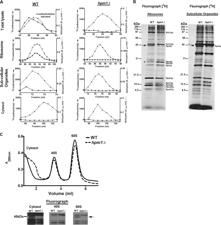
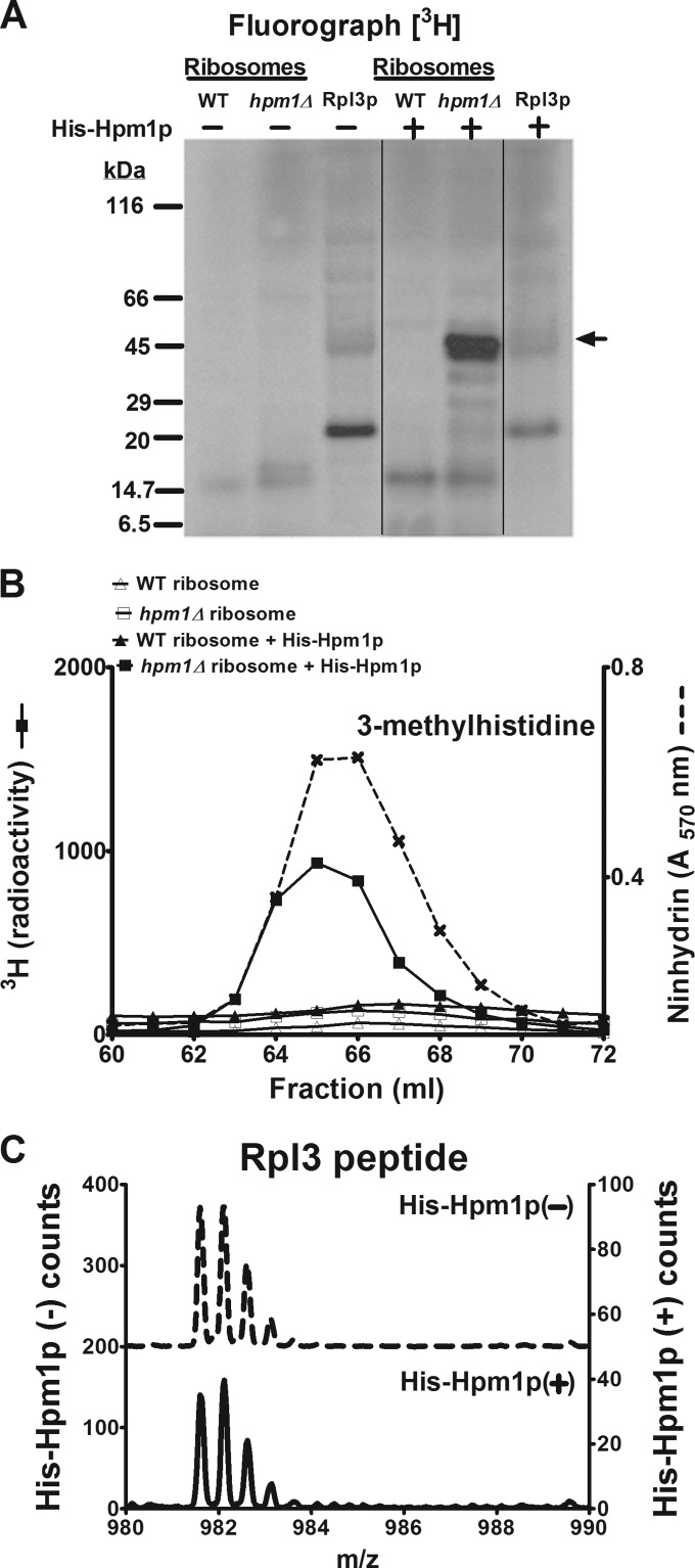
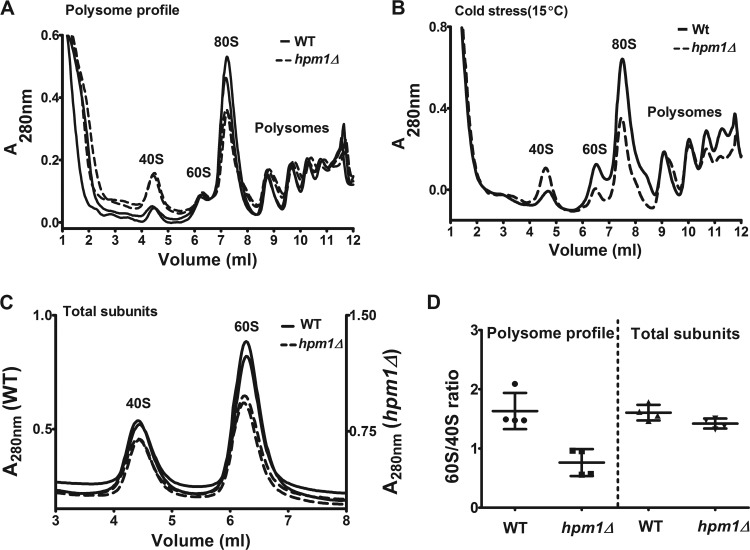
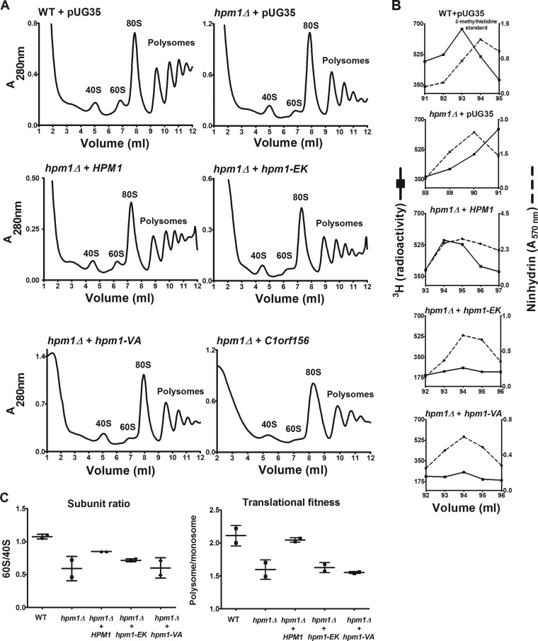
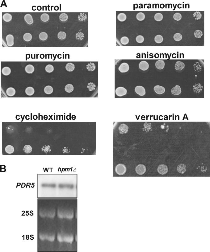
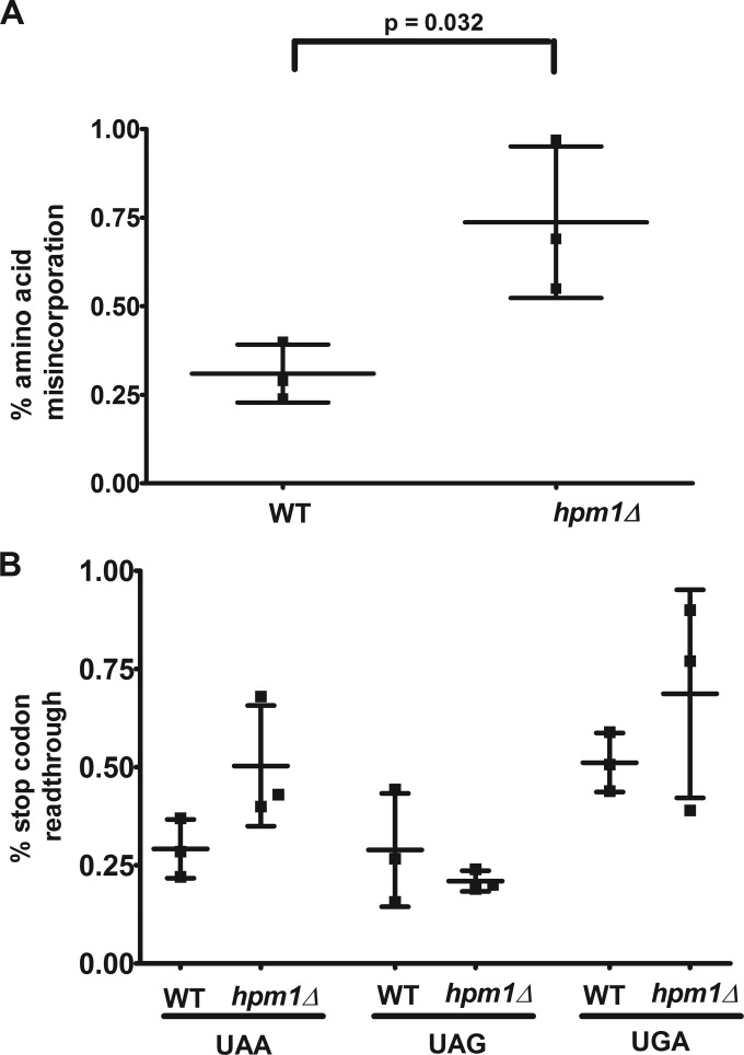
Histidine protein methylation is an unusual posttranslational modification. In the yeast Saccharomyces cerevisiae, the large ribosomal subunit protein Rpl3p is methylated at histidine 243, a residue that contacts the 25S rRNA near the P site. Rpl3p methylation is dependent upon the presence of Hpm1p, a candidate seven-beta-strand methyltransferase. In this study, we elucidated the biological activities of Hpm1p in vitro and in vivo. Amino acid analyses reveal that Hpm1p is responsible for all of the detectable protein histidine methylation in yeast. The modification is found on a polypeptide corresponding to the size of Rpl3p in ribosomes and in a nucleus-containing organelle fraction but was not detected in proteins of the ribosome-free cytosol fraction. In vitro assays demonstrate that Hpm1p has methyltransferase activity on ribosome-associated but not free Rpl3p, suggesting that its activity depends on interactions with ribosomal components. hpm1 null cells are defective in early rRNA processing, resulting in a deficiency of 60S subunits and translation initiation defects that are exacerbated in minimal medium. Cells lacking Hpm1p are resistant to cycloheximide and verrucarin A and have decreased translational fidelity. We propose that Hpm1p plays a role in the orchestration of the early assembly of the large ribosomal subunit and in faithful protein production.


Figures

FIG 1
FIG 1
Yeast protein 3-methylhistidine is found primarily on a ribosomal protein corresponding to Rpl3p and is dependent on HPM1. (A) Total lysate, ribosomes, subcellular organelles, and cytosol were prepared from WT (BY4742) and hpm1Δ mutant cells (BY4742) for amino acid analysis as described in Materials and Methods. The radioactivity shown (in counts per minute) has been normalized by background subtraction. Ninhydrin absorbance at 570 nm was measured to detect the 3-methylhistidine standard. (B) In vivo 3H-radiolabeled ribosomes and subcellular organelles from WT and hpm1Δ mutant cells (40 μg of protein) were pretreated with 2 U of Benzonase (Novagen 70746-4) for 30 min at 37°C and resolved by SDS-PAGE. 3H-methylated proteins were detected by fluorography for 13 weeks at −80°C as described in Materials and Methods. The arrow shows the methylated polypeptide corresponding to the molecular weight of Rpl3p, and the asterisk shows the positions of Rps2 and Rps3, whose levels of methylation are altered in the absence of HPM1. Radiolabeled bands corresponding to known methylated ribosomal proteins are identified on the right (14–22). (C) Seven OD units of WT and hpm1Δ mutant cells was radiolabeled with [3H]AdoMet, and the small 40S and large 60S ribosomal subunits were dissociated as described in Materials and Methods. One-hundred-microliter volumes of the cytosol, 40S, and 60S peak fractions were resolved by SDS-PAGE as described in Materials and Methods, except that a 12% Bis-Tris gel was prepared and run with MOPS running buffer. 3H-methylated proteins were detected by fluorography for 10 weeks at −80°C as described in Materials and Methods. At the bottom are three portions of lanes from the same gel.
FIG 2
FIG 2
Hpm1p can methylate Rpl3p on intact ribosomes but not free Rpl3p. (A) A 46-μg sample of crude ribosome protein isolated as described in Materials and Methods from WT and hpm1Δ mutant cells or 46 μg of recombinant Rpl3p was incubated with or without recombinant, histidine-tagged Hpm1p (His-Hpm1p; 12 μg) in the presence of 1 μM [3H]AdoMet and 100 mM sodium chloride–100 mM sodium phosphate (pH 7.5) (methylation buffer) for 5 h at 30°C. Reactions were terminated by the addition of equal volume of 2× Laemmli sample buffer, and proteins were analyzed by SDS-PAGE and fluorography as described in Materials and Methods. Film was incubated with the dried gel for 7 weeks at −80°C. The arrow indicates the approximate position of recombinant His-Rpl3p. The radiolabeled band at about 20 kDa appears to reflect a bacterial contaminant in the His-Rpl3p preparation. Vertical lines show where nonrelevant lanes were removed from the single gel. (B) Thirty-seven-microgram samples of crude ribosome protein from WT and hpm1Δ mutant cells were incubated with or without His-Hpm1p as described above. Proteins were trichloroacetic acid precipitated and acid hydrolyzed for amino acid analysis as described in Materials and Methods, except that fractions eluting at 60 to 72 ml were collected and the 3H radioactivity of 900 μl of each fraction was counted (reported in counts per minute). Ninhydrin, absorbance of the 3-methylhistidine standard. (C) The synthetic peptide WGTKKLPRKTHRGLRK (Biosynthesis, Lewisville, TX), corresponding to the methylated region of Rpl3p, was incubated with or without His-Hpm1p (30 μg) in the presence of 200 μM S-adenosyl-l-methionine p-toluenesulfonate (Sigma) and methylation buffer for 16 h at 30°C. Reactions were terminated with trifluoroacetic acid to a final concentration of 1%, and the products were fractionated by high-performance liquid chromatography with a PLRP-S reverse-phase column (pore size, 300 Å; bead size, 5 μm; 120 by 2 mm; Polymer Laboratories, Amherst, MA). The column was maintained at 50°C and initially equilibrated in 95% solvent A (0.1% trifluoroacetic acid in water) and 5% solvent B (0.1% trifluoroacetic acid in acetonitrile) at a flow rate of 0.5 ml/min. The following program was used: 10 min of 5% B, 25 min of a gradient to 60% B, 1 min of a gradient to 100% B, 5 min of 100% B, 1 min of a gradient to 5% B, and 8 min of 5% B. The column effluent was directed to the electrospray ion source of a QSTAR Elite (Applied Biosystems) mass spectrometer running in MS-only mode and was calibrated with external peptide standards. The left y axis represents the counts in the absence of His-Hpm1p, and the right y axis represents the counts in the presence of His-Hpm1p.
FIG 3
FIG 3
Lack of HPM1 results in deficiencies in large-subunit biogenesis and translation initiation. (A) Polysome profile analyses of WT and hpm1Δ mutant cells were done as described in Materials and Methods. Shown are two independent profiles each of the WT and of the hpm1Δ mutant. (B) Polysome profile analysis of cold-stressed (15°C) cells was done as described for panel A. (C) Total subunit analysis of WT and hpm1Δ mutant cells was done as described for panel A, except that cells were not pretreated with cycloheximide, the lysis buffer was replaced with buffer C, and 2 A260 units of extract was loaded. (D) Quantification of ratios of large to small ribosomal subunits for both the polysome profiles (A) and total subunits (C). Peak areas were determined with Graphical Analysis 3.8.4 software. Error bars represent standard deviations of four independent experiments. Unpaired t test two-tailed P values for the differences in the subunit ratios were 0.004 for the polysome ratio and 0.057 for the dissociated subunit ratio.
FIG 4
FIG 4
Loss of HPM1 results in early rRNA processing defects and delayed kinetics of pre-rRNA maturation. (A) rRNA precursor processing pathway in S. cerevisiae. The 35S pre-rRNA encodes the 18S rRNA of the small ribosomal subunit and the 5.8S and 25S rRNAs of the large subunit. The predominant pathway for the processing of the 35S pre-rRNA transcript is shown on the left, and the abnormal pathway is shown on the right (dashed arrow). First, the 5′ end of the 35S pre-rRNA is cleaved at sites A0 and A1, generating the mature 5′ end of the 18S rRNA. Cleavage at the A2 site separates the 20S and 27SA2 precursors. The 20S precursor is cleaved at site D in the cytoplasm to yield the mature 18S transcript. The 5′ end of the 27SA2 precursor is cleaved at site A3; this is followed by 5′-to-3′ exonuclease trimming to generate the mature 5′ end of the 5.8S rRNA. C2 cleavage separates the 5.8S and 25S precursors, which are then trimmed by exonucleases to yield the mature 5.8S and 25S rRNAs. (B) Northern blot analysis of rRNA precursor steady-state levels in the WT and the hpm1Δ mutant (lanes 1 and 2). Lanes 3 to 8 represent deletions of various ribosomal protein methyltransferases. The locations of the probes within each rRNA species are indicated on the right. The 25S and 18S blots are from the ethidium bromide fluorescence of the gel prior to transfer. (C) Top, Northern blot analysis of rRNA precursor steady-state levels with the 35S probe in WT and hpm1Δ and rnt1Δ mutant cells; bottom, 25S and 18S rRNAs detected by ethidium bromide fluorescence of the gel prior to transfer. (D) Top, pulse-chase assay with [3H]uracil for analysis of pre-rRNA processing kinetics in WT and hpm1Δ mutant cells. Cultures in the early exponential growth phase were pulsed with tritiated uracil for 2 min and then chased with an excess of nonisotopically labeled uracil. Bottom, analysis of the membrane from the pulse-chase assay by Northern blotting of 25S and 18S rRNAs. The ratios of the 25S to 18S rRNAs are normalized to the 1-min chase lane of the WT.
FIG 5
FIG 5
Plasmids containing WT HPM1 but not the human homolog C1orf156 can rescue hpm1Δ mutant defects in large-subunit biogenesis and translational initiation. (A) hpm1Δ mutant cells were transformed with the pUG35 plasmid containing the HPM1 gene, its human homolog C1orf156, or HPM1 active-site mutants under the control of the Met25 promoter. As controls, WT and hpm1Δ mutant cells were transformed with empty pUG35 expressing only GFP. The hpm1(EK) and hpm1(VA) genes have mutations in motif 1 of the MT domain, which is involved in AdoMet binding, i.e., EIGCG to KIVCE (EK) and VEIG to AAAA (VA). Cells were grown overnight in SD-Ura-Met at 30°C in a rotary shaker. Cells were then diluted in 100 ml of fresh SD-Ura-Met to an OD600 of 0.01 and grown overnight at 30°C until they reached an OD600 of 0.8 to 1.0. Polysome profile analysis was done as described in Materials and Methods. The HPM1, C1orf156, hpm1(EK), and hpm1(VA) genes were cloned into pUG35 with their stop codons to express the genes without the GFP tag. (B) WT and hpm1Δ mutant cells containing the plasmids described in panel A were labeled in vivo with [3H]AdoMet as described in Materials and Methods, except that 3.5 OD600 units of cells was labeled for 1 h. Total lysates were prepared in buffer C and acid hydrolyzed for amino acid analysis as described in Materials and Methods, except that the column was kept at 35°C and that 900 μl of each fraction was taken for radioactivity determination. 3H radioactivity is shown in counts per minute, and ninhydrin absorbance of the 3-methylhistidine standard was measured. (C) Quantification of polysome profiles. Ratios were calculated by determining the areas of the 40S, 60S, monosome (80S), and polysome peaks with Graphical Analysis 3.8.4 software. Error bars represent standard deviations of two independent experiments. The subunit ratio and translational fitness differences were not statistically significant at the P < 0.05 level.
FIG 6
FIG 6
Lack of HPM1 results in greatly increased resistance to the ribosome-targeting drugs cycloheximide and verrucarin A. (A) Dilution spot assays were done with mid-log-phase yeast cells (OD600 of 0.5) grown in YPD medium at 30°C in a rotary shaker for 2 generations from an overnight culture grown in YPD at 30°C. Cells were serially diluted 5-fold on YPD agar plates in the presence or absence of the ribosome-targeting antibiotics puromycin (50 μg/ml), cycloheximide (500 ng/ml), paromomycin (50 μg/ml), anisomycin (5 μg/ml), and verrucarin A (2 μg/ml) and incubated at 30°C for 2 days (control, puromycin, anisomycin, and paromomycin), 3 days (verrucarin A), or 5 days (cycloheximide). WT and hpm1Δ mutant cells containing the empty pUG35 vector were also spotted onto SD-Ura-Met and incubated for 3 days. (B) Northern blot analysis of mRNA expressed from the PDR5 gene encoding the multidrug exporter PDR5. The 25S and 18S blots are from the ethidium bromide fluorescence of the gel prior to transfer.
FIG 7
FIG 7
Cells deficient in Hpm1p have increased amino acid misincorporation and stop codon readthrough. The dual-luciferase assay was done as described in Materials and Methods. Percentages of amino acid misincorporation (A) and stop codon readthrough (B) were calculated by dividing the firefly/Renilla luciferase luminescence ratio by the same ratio of the respective control vector. Error bars represent the standard deviations of three independent experiments. The unpaired t test two-tailed P value is shown for panel A. Differences in stop codon readthrough between the WT and the hpm1Δ mutant were not statistically significant at the P < 0.05 level.

Similar articles

Cited by

References

    1. Lapeyre B. 2005. Conserved ribosomal RNA modification and their putative roles in ribosome biogenesis and translation, p 263–284 In Grosjean H. (ed), Fine-tuning of RNA functions by modification and editing. Springer, Berlin, Germany
    1. Johansson MO, Byström A. 2005. Transfer RNA modifications and modifying enzymes in Saccharomyces cerevisiae, p 87–120 In Grosjean H. (ed), Fine-tuning of RNA functions by modification and editing. Springer, Berlin, Germany
    1. Bokar J. 2005. The biosynthesis and functional roles of methylated nucleosides in eukaryotic mRNA, p 141–177 In Grosjean H. (ed), Fine-tuning of RNA functions by modification and editing. Springer, Berlin, Germany
    1. Polevoda B, Sherman F. 2007. Methylation of proteins involved in translation. Mol. Microbiol. 65:590–606. 10.1111/j.1365-2958.2007.05831.x - DOI - PubMed
    1. Katz JE, Dlakic M, Clarke SG. 2003. Automated identification of putative methyltransferases from genomic open reading frames. Mol. Cell. Proteomics 2:525–540 - PubMed

Publication types

MeSH terms