. 2014 Aug;34(8):1411-9.
doi: 10.1038/jcbfm.2014.101. Epub 2014 May 28.

Role of spleen-derived monocytes/macrophages in acute ischemic brain injury

Affiliations

Role of spleen-derived monocytes/macrophages in acute ischemic brain injury

Eunhee Kim et al. J Cereb Blood Flow Metab. 2014 Aug.

Abstract

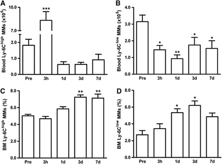
Monocytes/macrophages (MMs), mononuclear phagocytes, have been implicated in stroke-induced inflammation and injury. However, the presence of pro-inflammatory Ly-6C(high) and antiinflammatory Ly-6C(low) monocyte subsets raises uncertainty regarding their role in stroke pathologic assessment. With recent identification of the spleen as an immediate reservoir of MMs, this current study addresses whether the spleen-derived MMs are required for stroke pathologic assessment. We observed that the spleen was contracted in poststroke animals and the contraction was accompanied by decreased number of Ly-6C(high) and Ly-6C(low) subsets in the spleen. The deployment of these subsets from the spleen temporally coincided with respective increases in the ischemic brain. Compared to mice with the spleen, mice receiving a splenectomy just before the stroke displayed less accumulation of Ly-6C(high) and Ly-6C(low) MMs in the brain. Despite the reduced accumulation of both subsets, infarct size and swelling were not reduced in the asplenic mice. The dissociative findings of infarct size and extent of MM infiltration in the postischemic brain indicate minimal involvement of spleen-derived total MMs in acute infarct development. Selective Ly-6C(high) or Ly-6C(low) MM targeting is suggested to address the contribution of the individual subset to acute stroke pathologic assessment.


Figures

Figure 1
Figure 1
Stroke causes the reduction in spleen size and monocytes/macrophages (MMs). (A) Spleen weight before the stroke (pre), 3 hours, 1 day, 3 days, or 7 days after stroke. (B) Flow cytometry analyses of total leukocytes (R1) and total MMs (R2) in the spleen. R1 cells were further analyzed using a lineage marker (Lin) and CD11b antibodies to selectively gate the MM population (R2, Lin−/CD11b+). SSC, side scatter, index of cellular complexity; FSC, forward scatter, index of cell size. (C) Stroke-induced changes in total leukocytes (R1). (D) Stroke-induced changes in total MMs (R2). (E) Percentage (%) of MMs in total leukocytes, n=7 to 12/group, *P<0.05, **P<0.01, and ***P<0.001 versus pre; one-way analysis of variance. % Annexin V+ apoptotic cells in total leukocytes (F) and MMs (G) in the spleen at sham-stroke (Sham) or 1 day post stroke (1d-post). n=4 to 7/group, Student's t-test.
Figure 2
Figure 2
Stroke differentially reduces Ly-6Chigh and Ly-6Clow MM subsets in the spleen. The MM subsets were measured prior to ischemia (pre), 3 hours, 1 day, 3 days, or 7 days after stroke. (A) Flow cytometry analysis of MM subsets. Total MMs, indicated by R2, were further analyzed by Ly-6C expression. Cells without Ly-6C antibody serve as control (w/o antibody). Quantification of Ly-6Chigh (B) and Ly-6Clow subset (C) before and after stroke. n=7 to 12/group, **P<0.01, and ***P<0.001 versus pre. One-way analysis of variance. MMs, monocytes/macrophages.
Figure 3
Figure 3
Stroke-induced changes of MM subsets in the circulation and BM. MM subsets in the blood or BM were measured in mice prior to ischemia (pre, n=5) and 3 hours (n=11), 1 day (n=13), 3 days (n=10), or 7 days (n=6) post ischemia. Flow cytometric analyses using a lineage marker (Lin) and CD11b antibodies to selectively gate MM population (R2, Lin−/CD11b+ shown in Figure 1B) followed by Ly-6C antibody incubation. Ly-6Chigh (A) and Ly-6Clow (B) MM subsets in whole blood. Percentage of Ly-6Chigh (C) and Ly-6Clow MMs (D) from BM. *P<0.05, **P<0.01, and ***P<0.001 versus pre; one-way analysis of variance. BM, bone marrow; MMs, monocytes/macrophages.
Figure 4
Figure 4
Stroke causes the accumulation of peripheral MMs in the ipsilateral hemisphere. (A) Photomicrographs of GFP and CD11b immunostaining. Brain sections of C57BL/6 mice that received GFP+ splenic mononuclear cells 3 days after stroke and killed 6 hours after the cell infusion. GFP+ (a), CD11b+ (b), and GFP/CD11b colocalization in contralateral hemisphere (c) or peri-infarct area (d), GFP+/CD11b+ cells (e) or GFP+/CD11b− cells (f) of the peri-infarct area in high magnification, blue, DAPI-stained nucleus (d,e,f); scale bar=100 μm. (B) Representative flow cytometry plots in the contralateral and ipsilateral hemisphere at 3 day post ischemia shows two distinct populations: infiltrated MMs (CD45high/CD11b+) and resident microglia (CD45low/CD11b+). Dotted lines indicate antibody controls. (C) Quantification of total MMs in the ipsilateral hemisphere prior to ischemia (pre, n=5), 3 hours (n=7), 1 day (n=11), 3 days (n=9), or 7 days (n=6) post ischemia. *P<0.05, **P<0.01, and ***P<0.001 versus contralateral hemisphere, Student's t-test. GFP, green fluorescent protein; MMs, monocytes/macrophages.
Figure 5
Figure 5
Stroke causes the accumulation of both Ly-6Chigh and Ly-6Clow MMs in the brain. (A) Representative flow cytometry plots for two MM subsets by Ly-6C expression in the ipsilateral brain at 3 days post ischemia. R3, gate for infiltrated MMs. Dotted lines indicate antibody controls. Quantification of Ly-6Chigh (B) or Ly-6Clow MM subsets (C) prior to ischemia (pre, n=5), 3 hours (n=7), 1 day (n=11), 3 days (n=9), or 7 days (n=6) post ischemia. *P<0.05, **P<0.01, and ***P<0.001 versus contralateral hemisphere, Student's t-test. Mean Ly-6C fluorescence of Ly-6Chigh (D) and Ly-6Clow (E) subsets in the ipsilateral hemisphere (1, 3, and 7 days post). n=6 to 11/group (7 days post, n=6), ***P<0.001 versus 1 day post; one-way analysis of variance. MMs, monocytes/macrophages.
Figure 6
Figure 6
Splenectomy reduces the number of both MM subsets in the ischemic brain. Quantification of Ly-6Chigh (A) and Ly-6Clow (B) MMs in the brain of sham-splenectomized (Sham) or splenectomized (Spx) mice at 3 hours (n=4 to 7), 1 day (n=8 to 11), 3 days (n=7 to 8), and 7 days (n=6) post ischemia. *P<0.05, **P<0.01 versus sham; Student's t-test. MMs, monocytes/macrophages.
Figure 7
Figure 7
Spleen-derived MMs are not a major cause of stroke-induced injury. (AD) Infarct size and percentage of hemisphere swelling measured in sham-splenectomized (Sham) or splenectomized (Spx) mice at 3 days (A and B) and 7 days post ischemia (C and D). n=21 to 24/group (3 days post) or n=16 to 20/group (7 days post). Data expressed as mean±95% confidence interval. Student's t-test. MMs, monocytes/macrophages; NS, not significant.

References

    1. Gelderblom M, Leypoldt F, Steinbach K, Behrens D, Choe CU, Siler DA, et al. Temporal and spatial dynamics of cerebral immune cell accumulation in stroke. Stroke. 2009;40:1849–1857. - PubMed
    1. Jin R, Yang G, Li G. Inflammatory mechanisms in ischemic stroke: role of inflammatory cells. J Leukoc Biol. 2010;87:779–789. - PMC - PubMed
    1. Stevens SL, Bao J, Hollis J, Lessov NS, Clark WM, Stenzel-Poore MP. The use of flow cytometry to evaluate temporal changes in inflammatory cells following focal cerebral ischemia in mice. Brain Res. 2002;932:110–119. - PubMed
    1. Schilling M, Strecker JK, Schabitz WR, Ringelstein EB, Kiefer R. Effects of monocyte chemoattractant protein 1 on blood-borne cell recruitment after transient focal cerebral ischemia in mice. Neuroscience. 2009;161:806–812. - PubMed
    1. Tanaka R, Komine-Kobayashi M, Mochizuki H, Yamada M, Furuya T, Migita M, et al. Migration of enhanced green fluorescent protein expressing bone marrow-derived microglia/macrophage into the mouse brain following permanent focal ischemia. Neuroscience. 2003;117:531–539. - PubMed

Publication types

MeSH terms

LinkOut - more resources