Review
. 2014 Aug;52(8):713-37.
doi: 10.1002/dvg.22794. Epub 2014 Jun 24.

The functional diversity of essential genes required for mammalian cardiac development

Affiliations
Free PMC article
Review

The functional diversity of essential genes required for mammalian cardiac development

Christopher Clowes et al. Genesis. 2014 Aug.
Free PMC article

Abstract

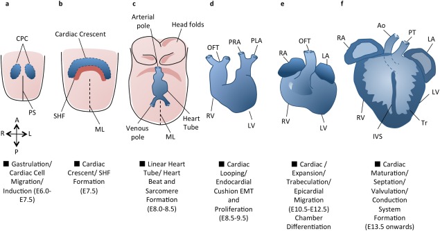
Genes required for an organism to develop to maturity (for which no other gene can compensate) are considered essential. The continuing functional annotation of the mouse genome has enabled the identification of many essential genes required for specific developmental processes including cardiac development. Patterns are now emerging regarding the functional nature of genes required at specific points throughout gestation. Essential genes required for development beyond cardiac progenitor cell migration and induction include a small and functionally homogenous group encoding transcription factors, ligands and receptors. Actions of core cardiogenic transcription factors from the Gata, Nkx, Mef, Hand, and Tbx families trigger a marked expansion in the functional diversity of essential genes from midgestation onwards. As the embryo grows in size and complexity, genes required to maintain a functional heartbeat and to provide muscular strength and regulate blood flow are well represented. These essential genes regulate further specialization and polarization of cell types along with proliferative, migratory, adhesive, contractile, and structural processes. The identification of patterns regarding the functional nature of essential genes across numerous developmental systems may aid prediction of further essential genes and those important to development and/or progression of disease.

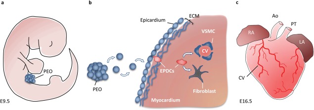
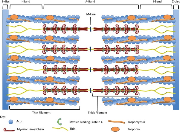
Keywords: cardiac chamber specification; epicardium; heart; heart fields; sarcomere; trabeculation.


Figures

Figure 1
Figure 1
An overview of cardiac development. Cardiac development progresses from the specification of cardiac progenitor cells (a) to the migration of these cells towards the midline to form the cardiac crescent (b). The developing heart then forms a linear tube (c), which undergoes dextral looping to acquire the appropriate left–right asymmetry (d). The heart tube is further subdivided into the four chambers (e), and the maturation of the endocardial cushions into the valves and development of the great vessels provides for unidirectional blood flow through the chambers (f). Adapted from (Buckingham et al., 2005). A = Anterior, Ao = Aorta, CPC = Cardiac Precursor Cells, IVS = Interventricular septum, L = Left, LA = Left Atrium, LV = Left Ventricle, ML = Midline, OFT = Outflow Tract, P = Posterior, PHF = Primary Heart Field, PLA = Primitive Left Atrium, PRA = Primitive Right Atrium, PS = Primitive Streak, PT = Pulmonary Trunk, R = Right, RA = Right Atrium, RV = Right Ventricle, SHF = Secondary Heart Field, Tr = Trabeculae.
Figure 2
Figure 2
Ventricular trabeculation and endocardial cushion development. Schematic representation of the E9.5 looping mouse heart (a) with location of the outflow tract (left) and atrioventricular (right) endocardial cushions (ECC). Two different cross-sections through the E10.5 mouse heart (b) showing the midgestational location of the OFT cushions (left) and atrioventricular cushions (right). AVC = Atrioventricular canal, CA = Common atrium, IVS = Interventricular Septum, LA = Left Atrium, LV = Left Ventricle, OFT - Outflow Tract, RA = Right Atrium, RV = Right Ventricle, Tr = Trabeculae.
Figure 3
Figure 3
The PEO contributes to CV formation. (a) The proepicardial organ (PEO) is a transient structure that forms near the sinus venosus of the postlooped embryonic heart at approximately E9.5. (b) Cells within the proepicardial organ translocate across the pericardial cavity and adhere to the myocardium to form an epithelial sheet that envelopes the developing heart, called the epicardium, by E11.0. Later in development, a sub population of epicardial cells undergo epithelial to mesenchymal transition, allowing them to migrate through the subepicardial ECM and invade the myocardium. These EPDCs differentiate into VSMC and cardiac fibroblasts, and are essential for the formation of the mature CV, as observed on the ventricular surface of the E16.5 heart (c). Ao = Aorta, LA = Left Atria, PT = Pulmonary Trunk, RA = Right Atria.
Figure 4
Figure 4
Sarcomere structure. Schematic representation of the mouse sarcomere showing the relevant banding pattern according to its electron microscopic appearance. Thin filaments are comprised of two interacting helically organized F-Actin polymeric chains along which Tropomyosin polymers lie in the grooves. Troponin complexes are found at set periods along the thin filament. Thick filaments are comprised of over 300 Myosin II molecules arranged into filament bundles and many interacting nonmyosin proteins. Individual molecules of the giant protein Titin span from Z-disc to M-line. Adapted from (Morimoto, 2008).

Similar articles

Cited by

References

    1. Aanhaanen WT, Boukens BJ, Sizarov A, Wakker V, de Gier-de Vries C, van Ginneken AC, Moorman AF, Coronel R, Christoffels VM. Defective Tbx2-dependent patterning of the atrioventricular canal myocardium causes accessory pathway formation in mice. J Clin Invest. 2011;121:534–544. - PMC - PubMed
    1. Aanhaanen WT, Brons JF, Dominguez JN, Rana MS, Norden J, Airik R, Wakker V, de Gier-de Vries C, Brown NA, Kispert A, Moorman AF, Christoffels VM. The Tbx2+ primary myocardium of the atrioventricular canal forms the atrioventricular node and the base of the left ventricle. Circ Res. 2009;104:1267–1274. - PubMed
    1. Aanhaanen WT, Mommersteeg MT, Norden J, Wakker V, de Gier-de Vries C, Anderson RH, Kispert A, Moorman AF, Christoffels VM. Developmental origin, growth, and three-dimensional architecture of the atrioventricular conduction axis of the mouse heart. Circ Res. 2010;107:728–736. - PubMed
    1. Acharya A, Baek ST, Huang G, Eskiocak B, Goetsch S, Sung CY, Banfi S, Sauer MF, Olsen GS, Duffield JS, Olson EN, Tallquist MD. The bHLH transcription factor Tcf21 is required for lineage-specific EMT of cardiac fibroblast progenitors. Development. 2012;139:2139–2149. - PMC - PubMed
    1. Arai A, Yamamoto K, Toyama J. Murine cardiac progenitor cells require visceral embryonic endoderm and primitive streak for terminal differentiation. Dev Dyn. 1997;210:344–353. - PubMed

Publication types

LinkOut - more resources