Characterization of the expression and inflammatory activity of NADPH oxidase after spinal cord injury
- PMID: 24866054
- PMCID: PMC4183135
- DOI: 10.3109/10715762.2014.927578
Characterization of the expression and inflammatory activity of NADPH oxidase after spinal cord injury
Abstract
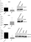
Reactive oxygen species (ROS) and the NADPH oxidase (NOX) enzyme are both up-regulated after spinal cord injury (SCI) and play significant roles in promoting post-injury inflammation. However, the cellular and temporal expression profile of NOX isotypes, including NOX2, 3, and 4, after SCI is currently unclear. The purpose of this study was to resolve this expression profile and examine the effect of inhibition of NOX on inflammation after SCI. Briefly, adult male rats were subjected to moderate contusion SCI. Double immunofluorescence for NOX isotypes and CNS cellular types was performed at 24 h, 7 days, and 28 days post-injury. NOX isotypes were found to be expressed in neurons, astrocytes, and microglia, and this expression was dependent on injury status. NOX2 and 4 were found in all cell types assessed, while NOX3 was positively identified in neurons only. NOX2 was the most responsive to injury, increasing in both microglia and astrocytes. The biggest increases in expression were observed at 7 days post-injury and increased expression was maintained through 28 days. NOX2 inhibition by systemic administration of gp91ds-tat at 15 min, 6 h or 7 days after injury reduced both pro-inflammatory cytokine expression and evidence of oxidative stress in the injured spinal cord. This study therefore illustrates the regional and temporal influence on NOX isotype expression and the importance of NOX activation in SCI. This information will be useful in future studies of understanding ROS production after injury and therapeutic potentials.
Keywords: NOX2; animal studies; cytokines; gp91ds-tat; oxidative stress.
Conflict of interest statement
No competing financial interests exist.
Figures


Similar articles
-
Cellular and temporal expression of NADPH oxidase (NOX) isotypes after brain injury.J Neuroinflammation. 2013 Dec 17;10:155. doi: 10.1186/1742-2094-10-155. J Neuroinflammation. 2013. PMID: 24344836 Free PMC article.
-
Inhibition of NOX2 reduces locomotor impairment, inflammation, and oxidative stress after spinal cord injury.J Neuroinflammation. 2015 Sep 17;12:172. doi: 10.1186/s12974-015-0391-8. J Neuroinflammation. 2015. PMID: 26377802 Free PMC article.
-
Differential effects of NOX2 and NOX4 inhibition after rodent spinal cord injury.PLoS One. 2023 Mar 10;18(3):e0281045. doi: 10.1371/journal.pone.0281045. eCollection 2023. PLoS One. 2023. PMID: 36897852 Free PMC article.
-
Central Nervous System Injury and Nicotinamide Adenine Dinucleotide Phosphate Oxidase: Oxidative Stress and Therapeutic Targets.J Neurotrauma. 2017 Feb 15;34(4):755-764. doi: 10.1089/neu.2016.4486. Epub 2016 Jun 27. J Neurotrauma. 2017. PMID: 27267366 Free PMC article. Review.
-
New insights on NOX enzymes in the central nervous system.Antioxid Redox Signal. 2014 Jun 10;20(17):2815-37. doi: 10.1089/ars.2013.5703. Epub 2014 Jan 16. Antioxid Redox Signal. 2014. PMID: 24206089 Free PMC article. Review.
Cited by
-
Green Synthesis, Chemical Characterization, and Assessment of the Neuroprotective Properties of Zinc Nanoparticles on the Spinal Cord Injury Contusive Model in Rats.Biol Trace Elem Res. 2025 Aug 7. doi: 10.1007/s12011-025-04766-z. Online ahead of print. Biol Trace Elem Res. 2025. PMID: 40773054
-
NADPH-Oxidase 2 Promotes Autophagy in Spinal Neurons During the Development of Morphine Tolerance.Neurochem Res. 2021 Aug;46(8):2089-2096. doi: 10.1007/s11064-021-03347-5. Epub 2021 May 18. Neurochem Res. 2021. PMID: 34008119
-
NOX2 is involved in CB2-mediated protection against lung ischemia-reperfusion injury in mice.Int J Clin Exp Pathol. 2020 Feb 1;13(2):277-285. eCollection 2020. Int J Clin Exp Pathol. 2020. PMID: 32211110 Free PMC article.
-
A Novel Rac1-GSPT1 Signaling Pathway Controls Astrogliosis Following Central Nervous System Injury.J Biol Chem. 2017 Jan 27;292(4):1240-1250. doi: 10.1074/jbc.M116.748871. Epub 2016 Dec 9. J Biol Chem. 2017. PMID: 27941025 Free PMC article.
-
Neuronal NADPH oxidase 2 regulates growth cone guidance downstream of slit2/robo2.Dev Neurobiol. 2021 Jan;81(1):3-21. doi: 10.1002/dneu.22791. Epub 2020 Dec 5. Dev Neurobiol. 2021. PMID: 33191581 Free PMC article.
References
-
- Faul M, Xu L, Wald MM, Coronado VG. Traumatic brain injury in the United States: emergency department visits, hospitalizations, and deaths. Centers for Disease Control and Prevention; 2010.
-
-
Center TNSS. Spinal cord injury: Facts and Figures. 2009.
-
-
- Gilgun-Sherki Y, Melamed E, Offen D. Oxidative stress induced-neurodegenerative diseases: the need for antioxidants that penetrate the blood brain barrier. Neuropharmacology. 2001;40:959–75. - PubMed
-
- Quinn MT, Ammons MC, Deleo FR. The expanding role of NADPH oxidases in health and disease: no longer just agents of death and destruction. Clin Sci (Lond) 2006;111:1–20. - PubMed
-
- Cave AC, Brewer AC, Narayanapanicker A, Ray R, Grieve DJ, Walker S, Shah AM. NADPH oxidases in cardiovascular health and disease. Antioxid Redox Signal. 2006;8:691–728. - PubMed
Publication types
MeSH terms
Substances
Grants and funding
LinkOut - more resources
Full Text Sources
Other Literature Sources
Medical
Miscellaneous
