. 2014 Jul;137(Pt 7):2040-51.
doi: 10.1093/brain/awu120. Epub 2014 May 27.

Sequestration of multiple RNA recognition motif-containing proteins by C9orf72 repeat expansions

Affiliations

Sequestration of multiple RNA recognition motif-containing proteins by C9orf72 repeat expansions

Johnathan Cooper-Knock et al. Brain. 2014 Jul.

Abstract

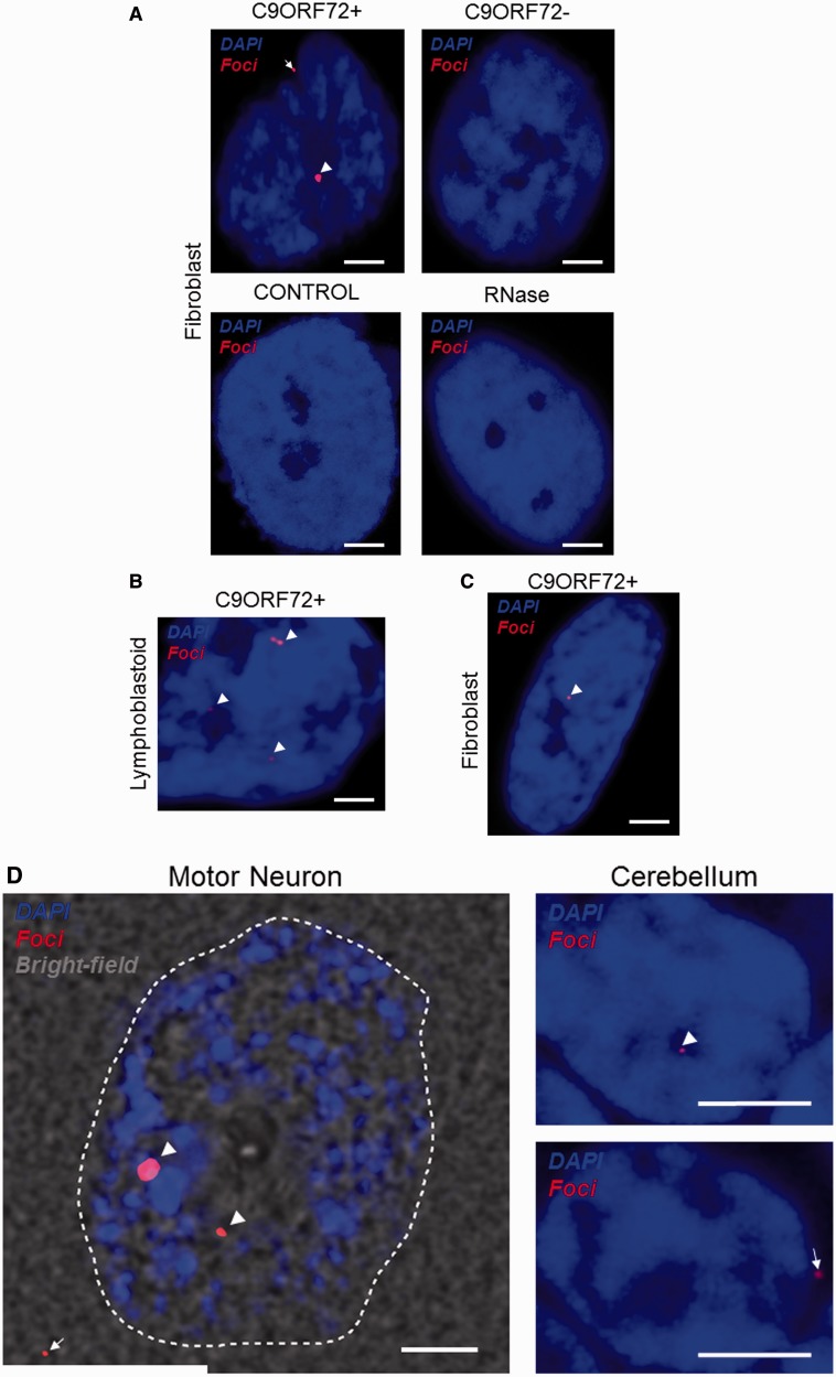
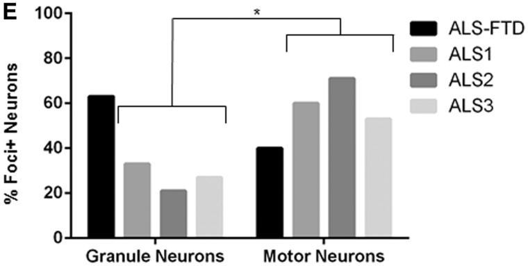
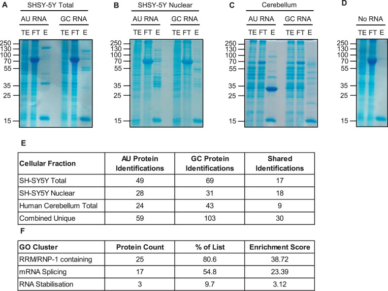
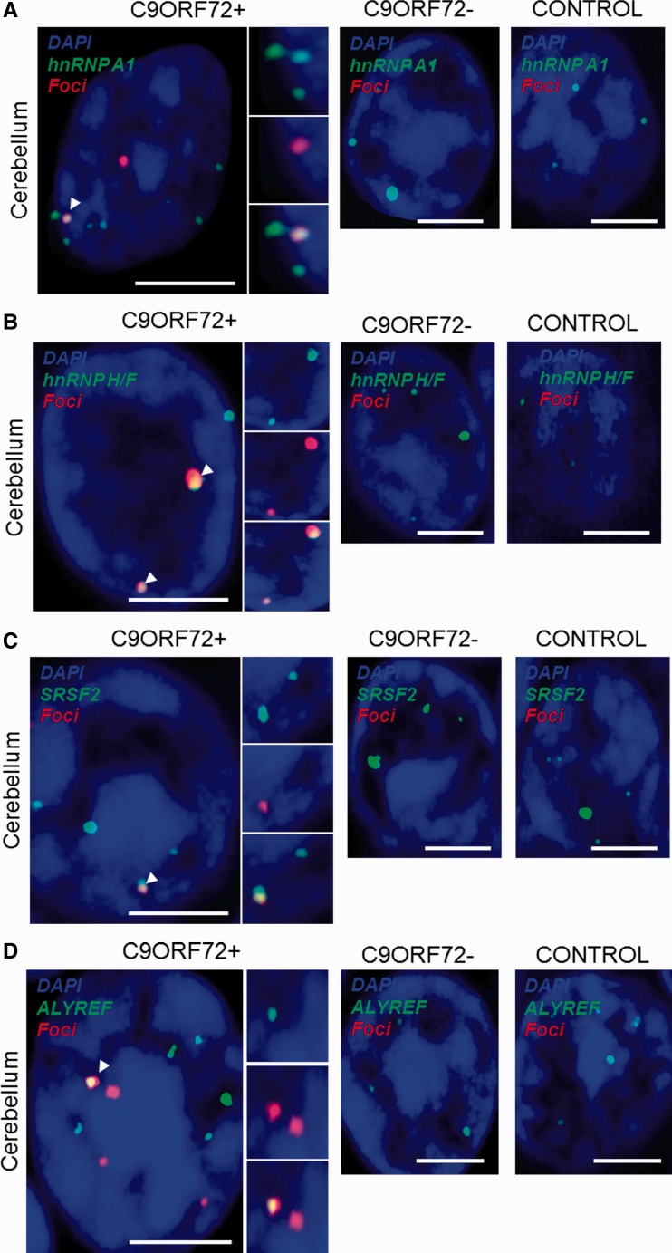
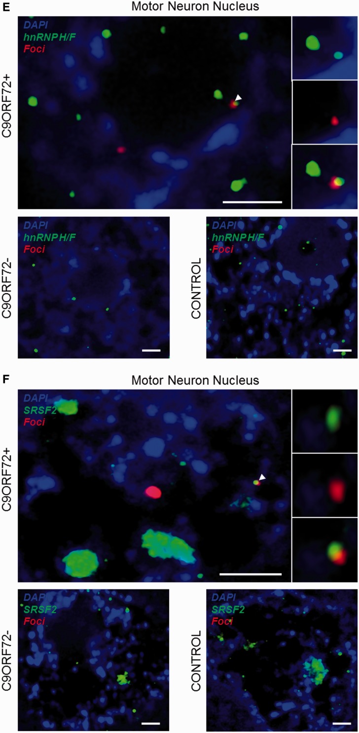
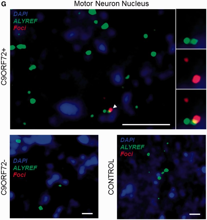
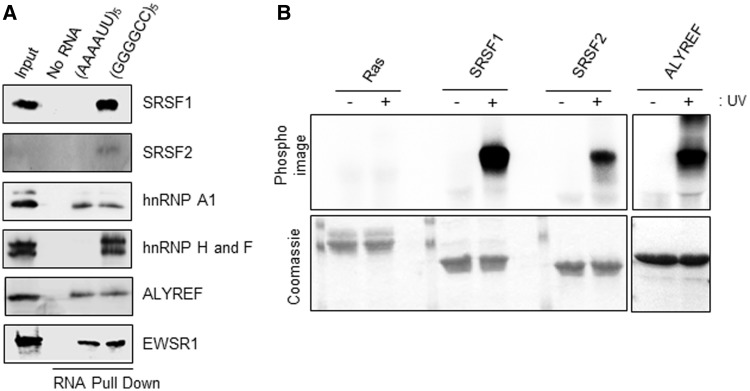
GGGGCC repeat expansions of C9orf72 represent the most common genetic variant of amyotrophic lateral sclerosis and frontotemporal degeneration, but the mechanism of pathogenesis is unclear. Recent reports have suggested that the transcribed repeat might form toxic RNA foci that sequester various RNA processing proteins. Consensus as to the identity of the binding partners is missing and whole neuronal proteome investigation is needed. Using RNA fluorescence in situ hybridization we first identified nuclear and cytoplasmic RNA foci in peripheral and central nervous system biosamples from patients with amyotrophic lateral sclerosis with a repeat expansion of C9orf72 (C9orf72+), but not from those patients without a repeat expansion of C9orf72 (C9orf72-) or control subjects. Moreover, in the cases examined, the distribution of foci-positive neurons correlated with the clinical phenotype (t-test P < 0.05). As expected, RNA foci are ablated by RNase treatment. Interestingly, we identified foci in fibroblasts from an asymptomatic C9orf72+ carrier. We next performed pulldown assays, with GGGGCC5, in conjunction with mass spectrometry analysis, to identify candidate binding partners of the GGGGCC repeat expansion. Proteins containing RNA recognition motifs and involved in splicing, messenger RNA nuclear export and/or translation were significantly enriched. Immunohistochemistry in central nervous system tissue from C9orf72+ patients with amyotrophic lateral sclerosis demonstrated co-localization of RNA foci with SRSF2, hnRNP H1/F, ALYREF and hnRNP A1 in cerebellar granule cells and with SRSF2, hnRNP H1/F and ALYREF in motor neurons, the primary target of pathology in amyotrophic lateral sclerosis. Direct binding of proteins to GGGGCC repeat RNA was confirmed in vitro by ultraviolet-crosslinking assays. Co-localization was only detected in a small proportion of RNA foci, suggesting dynamic sequestration rather than irreversible binding. Additional immunohistochemistry demonstrated that neurons with and without RNA foci were equally likely to show nuclear depletion of TDP-43 (χ(2) P = 0.75) or poly-GA dipeptide repeat protein inclusions (χ(2) P = 0.46). Our findings suggest two non-exclusive pathogenic mechanisms: (i) functional depletion of RNA-processing proteins resulting in disruption of messenger RNA splicing; and (ii) licensing of expanded C9orf72 pre-messenger RNA for nuclear export by inappropriate association with messenger RNA export adaptor protein(s) leading to cytoplasmic repeat associated non-ATG translation and formation of potentially toxic dipeptide repeat protein.

Keywords: amyotrophic lateral sclerosis; fluorescence imaging; genetics; pathology.


Figures

Figure 1
Figure 1
RNA FISH shows GGGGCC expanded RNA foci are found in peripheral cells and CNS tissue from C9orf72+ patients but not from C9orf72− ALS cases or controls. RNA foci (arrowheads) are present in fibroblasts (A), lymphoblastoid cells (B) and CNS tissue (D) from C9orf72+ patients with ALS, and in fibroblasts from a C9orf72+ asymptomatic carrier (C). RNA foci are ablated by RNase treatment (A). RNA foci are predominantly nuclear but cytoplasmic foci are observed in peripheral cells and CNS tissue (A and D, arrows). Abundance of foci in cerebellar granule cells and motor neurons has been quantified (E), in those cases where the initial clinical presentation was ALS the proportion of foci+ motor neurons is significantly higher (*P < 0.05). Scale bar = 3 µm. FTD = frontotemporal degeneration.
Figure 1
Figure 1
RNA FISH shows GGGGCC expanded RNA foci are found in peripheral cells and CNS tissue from C9orf72+ patients but not from C9orf72− ALS cases or controls. RNA foci (arrowheads) are present in fibroblasts (A), lymphoblastoid cells (B) and CNS tissue (D) from C9orf72+ patients with ALS, and in fibroblasts from a C9orf72+ asymptomatic carrier (C). RNA foci are ablated by RNase treatment (A). RNA foci are predominantly nuclear but cytoplasmic foci are observed in peripheral cells and CNS tissue (A and D, arrows). Abundance of foci in cerebellar granule cells and motor neurons has been quantified (E), in those cases where the initial clinical presentation was ALS the proportion of foci+ motor neurons is significantly higher (*P < 0.05). Scale bar = 3 µm. FTD = frontotemporal degeneration.
Figure 2
Figure 2
5’-[AAAAUU]5- and 5’-[GGGGCC]5 RNAs sequester distinct sets of proteins from human neuronal cell line fractions and dissected human cerebellar tissue. Pulldown assays using biotinylated RNAs (no RNA, 5’-[AAAAUU]5- or 5’-[GGGGCC]5) and extracts from total or nuclear fractions of SH-SY5Y cells, or human cerebellar tissue; I = input (1%); FT = flow through (1%); E = eluted (25%) (A–D). Mass spectrometry (MS) analysis of proteins co-purified with biotinylated RNAs (E). Gene ontology (GO) enrichment of SH-SY5Y nuclear hits (F).
Figure 3
Figure 3
Combined RNA FISH and immunohistochemistry demonstrate co-localization of nuclear speckle components with RNA foci in CNS tissue. hnRNP A1 (A), hnRNP H1/F (B), SRSF2 (C) and ALYREF (D) are observed to co-localize with RNA foci (arrowheads) in cerebellar granule cells from C9orf72+ patients with ALS. hnRNP H1/F (E), SRSF2 (F) and ALYREF (G) are observed to co-localize with RNA foci (arrowheads) in nuclei of motor neurons from C9orf72+ patients with ALS. Co-localization events are enlarged and unmerged protein and RNA foci are shown for comparison. The normal staining pattern of the two proteins in C9orf72− cases with ALS and control subjects is included for comparison. Scale bar = 3 µm.
Figure 3
Figure 3
Combined RNA FISH and immunohistochemistry demonstrate co-localization of nuclear speckle components with RNA foci in CNS tissue. hnRNP A1 (A), hnRNP H1/F (B), SRSF2 (C) and ALYREF (D) are observed to co-localize with RNA foci (arrowheads) in cerebellar granule cells from C9orf72+ patients with ALS. hnRNP H1/F (E), SRSF2 (F) and ALYREF (G) are observed to co-localize with RNA foci (arrowheads) in nuclei of motor neurons from C9orf72+ patients with ALS. Co-localization events are enlarged and unmerged protein and RNA foci are shown for comparison. The normal staining pattern of the two proteins in C9orf72− cases with ALS and control subjects is included for comparison. Scale bar = 3 µm.
Figure 4
Figure 4
Identified RNA-binding candidates interact specifically and directly with GGGGCC5 RNA. (A) Neuronal SH-SY5Y whole cell extract was incubated with either no RNA, AU-rich or GC-rich biotinylated RNA coated onto streptavidin beads before UV-cross linking. Bound proteins were eluted using RNase A and further identified using SDS-PAGE and western immunoblotting with the indicated antibodies. It is noted that the weak signal for SRSF2 is due to difficulty finding an antibody that is efficacious in western immunoblotting. The anti-hnRNP H1/F antibody recognizes both proteins, which are similar (Garneau et al., 2005). (B) Hexa-histidine-tagged recombinant SRSF1 11-196, GB1-tagged SRSF2 9-101 and ALYREF full length were expressed in E. coli and purified using metal ion affinity chromatography in 1 M NaCl containing buffers to remove potentially bound RNA from E. coli (bottom). GGGGCC5 RNA was separately end-labelled with poly nucleotide kinase using [γ-32P]-ATP, before incubation with purified proteins. RNA was covalently bound (+) or not (−) after UV irradiation. Absence of radioactive signal (top; PhosphoImage) in absence of UV irradiation demonstrates specificity of direct binding observed after UV treatment. All gels shown in the different panels were exposed simultaneously for the same amount of time (5 h).

References

    1. Ash PE, Bieniek KF, Gendron TF, Caulfield T, Lin WL, Dejesus-Hernandez M, et al. Unconventional translation of C9ORF72 GGGGCC expansion generates insoluble polypeptides specific to c9FTD/ALS. Neuron. 2013;77:639–46. - PMC - PubMed
    1. Bengoechea R, Tapia O, Casafont I, Berciano J, Lafarga M, Berciano MT. Nuclear speckles are involved in nuclear aggregation of PABPN1 and in the pathophysiology of oculopharyngeal muscular dystrophy. Neurobiol Dis. 2012;46:118–29. - PubMed
    1. Brettschneider J, Del Tredici K, Toledo JB, Robinson JL, Irwin DJ, Grossman M, et al. Stages of pTDP-43 pathology in amyotrophic lateral sclerosis. Ann Neurol. 2013;74:20–38. - PMC - PubMed
    1. Byrne S, Heverin M, Elamin M, Walsh C, Hardiman O. Intermediate repeat expansion length in C9orf72 may be pathological in amyotrophic lateral sclerosis. Amyotroph Lateral Scler Frontotemporal Degener. 2013;15:148–50. - PubMed
    1. Casafont I, Bengoechea R, Tapia O, Berciano MT, Lafarga M. TDP-43 localizes in mRNA transcription and processing sites in mammalian neurons. J Struct Biol. 2009;167:235–41. - PubMed

Publication types

MeSH terms