. 2014 Aug 15;9(8):1718-25.
doi: 10.1021/cb5002526. Epub 2014 Jun 6.

Substrate specificity of the lanthipeptide peptidase ElxP and the oxidoreductase ElxO

Affiliations

Substrate specificity of the lanthipeptide peptidase ElxP and the oxidoreductase ElxO

Manuel A Ortega et al. ACS Chem Biol. .

Abstract

The final step in lanthipeptide biosynthesis involves the proteolytic removal of an N-terminal leader peptide. In the class I lanthipeptide epilancin 15X, this step is performed by the subtilisin-like serine peptidase ElxP. Bioinformatic, kinetic, and mass spectrometric analysis revealed that ElxP recognizes the stretch of amino acids DLNPQS located near the proteolytic cleavage site of its substrate, ElxA. When the ElxP recognition motif was inserted into the noncognate lanthipeptide precursor NisA, ElxP was able to proteolytically remove the leader peptide from NisA. Proteolytic removal of the leader peptide by ElxP during the biosynthesis of epilancin 15X exposes an N-terminal dehydroalanine on the core peptide of ElxA that hydrolyzes to a pyruvyl group. The short-chain dehydrogenase ElxO reduces the pyruvyl group to a lactyl moiety in the final step of epilancin 15X maturation. Using synthetic peptides, we also investigated the substrate specificity of ElxO and determined the 1.85 Å resolution X-ray crystal structure of the enzyme.


Figures

Figure 1
Figure 1
(a) Biosynthesis of epilancin 15X, involving dehydration of Ser and Thr residues by ElxB to yield dehydroalanine (Dha, green) and dehydrobutyrine (Dhb, purple), formation of lanthionine (red) or methyllanthionine (blue) rings catalyzed by ElxC, removal of the leader peptide by the peptidase ElxP, and (b) reduction of the N-terminal pyruvyl moiety catalyzed by ElxO. Abu (2-aminobutyric acid), Pyr (pyruvyl), Lac (lactyl). The leader peptide is shown in bold font.
Figure 2
Figure 2
(a) Sequence alignment of selected LanA leader peptides for which the final products (class I lanthipeptides) have been structurally characterized. The FxLx motif is highlighted in green. The putative LanP recognition motifs are shown in blue and red boxes for the NisA-group and the ElxA-group, respectively. LanP cleavage sites are shown with an arrow. (b) MCMC phylogenetic tree of LanP enzymes corresponding to the LanA substrates shown in part a. Bayesian inferences of posterior probabilities are shown above or below the branches. Two LanPs involved in class II lanthipeptide biosynthesis (LicP for lichenicidin and CylA for cytolysin) served as the out group of the tree.
Figure 3
Figure 3
Kinetic characterization of ElxP peptidase activity for His6-ElxA and mutant peptides. Purified (a) ElxA, (b) ElxA Q−1A, (c) ElxA L−4A, and (d) ElxA P−2A were digested with MBP-ElxP and leader peptide formation was monitored at different time points by HPLC. The rate of MBP-ElxP catalysis was plotted as a function of different substrate concentrations. The data was fit to the Michaelis–Menten equation to give the kinetic parameters shown, presented as the average ± standard error of two independent experiments.
Figure 4
Figure 4
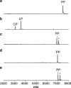
MALDI-TOF MS data on cleavage of NisA and NisA mutants by ElxP. (a) NisA (m/z 7992) treated with ElxP, (b) NisA-G−5D/A−4L/S−3N/R−1Q/Q−1_I1insA treated with ElxP, (c) NisA-R−1Q (m/z 7412) treated with ElxP, (d) NisA-R−1Q/Q−1_I1insA (m/z 7485) treated with ElxP, and (e) NisA-G−5D/A−4L/S−3N/R−1Q (m/z 7542) treated with ElxP. PP-precursor peptide, CP-core peptide, and LP-leader peptide. His6-NisA-G−5D/A−4L/S−3N/R−1Q/Q−1_I1insA unmodified core peptide, m/z 3568; leader peptide, m/z 4064. *Ion corresponding to peptide with gluconoylation of the His6-tag of NisA.
Figure 5
Figure 5
Schematic structure of lactocin S and formation of dihydrolactocin S. (a) The lantibiotic lactocin S contains an N-terminal Pyr group. (b) Lactocin S was converted to dihydrolactocin S. Left panel: MS analysis of lactocin S (calculated m/z = 3762.8851) incubated with NADPH in the absence of His6-ElxO. The peak at m/z = 3794.9348 corresponds to oxidized lactocin S (M+O). Right panel: MS analysis of dihydrolactocin S (calculated m/z = 3764.8851) generated after incubation of lactocin S with both NADPH and His6-ElxO. The peaks at m/z = 3786.8765 and 3808.8447 correspond to the sodium and disodium adducts of dihydrolactocin S.
Figure 6
Figure 6
(a) Single concentration and (b) serial dilution agar diffusion bioactivity assays. The samples spotted were enzymatically synthesized dihydrolactocin S (sample 1) and control samples lacking enzyme (sample 2), cofactor (sample 3), or both (sample 4) and incubated under the same reaction conditions. Sample 5 was a control assay lacking lactocin S. See also Supporting Information Figure 6.
Figure 7
Figure 7
(a) X-ray crystal structure of ElxO bound to NADPH. (b) Side chain residues important for binding of NADPH and catalysis in ElxO. c) ElxO surface representation depicting groove, which might serve as the putative peptide-binding site.

Similar articles

Cited by

References

    1. Arnison P. G.; Bibb M. J.; Bierbaum G.; Bowers A. A.; Bugni T. S.; Bulaj G.; Camarero J. A.; Campopiano D. J.; Challis G. L.; Clardy J.; Cotter P. D.; Craik D. J.; Dawson M.; Dittmann E.; Donadio S.; Dorrestein P. C.; Entian K.-D.; Fischbach M. A.; Garavelli J. S.; Göransson U.; Gruber C. W.; Haft D. H.; Hemscheidt T. K.; Hertweck C.; Hill C.; Horswill A. R.; Jaspars M.; Kelly W. L.; Klinman J. P.; Kuipers O. P.; Link A. J.; Liu W.; Marahiel M. A.; Mitchell D. A.; Moll G. N.; Moore B. S.; Müller R.; Nair S. K.; Nes I. F.; Norris G. E.; Olivera B. M.; Onaka H.; Patchett M. L.; Piel J.; Reaney M. J. T.; Rebuffat S.; Ross R. P.; Sahl H.-G.; Schmidt E. W.; Selsted M. E.; Severinov K.; Shen B.; Sivonen K.; Smith L.; Stein T.; Süssmuth R. E.; Tagg J. R.; Tang G.-L.; Truman A. W.; Vederas J. C.; Walsh C. T.; Walton J. D.; Wenzel S. C.; Willey J. M.; van der Donk W. A. (2013) Ribosomally synthesized and post-translationally modified peptide natural products: Overview and recommendations for a universal nomenclature. Nat. Prod. Rep. 30, 108–160. - PMC - PubMed
    1. Schnell N.; Entian K.-D.; Schneider U.; Götz F.; Zahner H.; Kellner R.; Jung G. (1988) Prepeptide sequence of epidermin, a ribosomally synthesized antibiotic with four sulphide-rings. Nature 333, 276–278. - PubMed
    1. van der Meer J. R.; Rollema H. S.; Siezen R. J.; Beerthuyzen M. M.; Kuipers O. P.; de Vos W. M. (1994) Influence of amino acid substitutions in the nisin leader peptide on biosynthesis and secretion of nisin by Lactococcus lactis. J. Biol. Chem. 269, 3555–3562. - PubMed
    1. Li B.; Yu J. P.; Brunzelle J. S.; Moll G. N.; van der Donk W. A.; Nair S. K. (2006) Structure and mechanism of the lantibiotic cyclase involved in nisin biosynthesis. Science 311, 1464–1467. - PubMed
    1. McClerren A. L.; Cooper L. E.; Quan C.; Thomas P. M.; Kelleher N. L.; van der Donk W. A. (2006) Discovery and in vitro biosynthesis of haloduracin, a two-component lantibiotic. Proc. Natl. Acad. Sci. U.S.A. 103, 17243–17248. - PMC - PubMed

Publication types