

Parameter comparison of white matter diffusion tensor imaging (DTI) in rhesus macaques (Macaca mulatta)
- PMID: 24866488
- PMCID: PMC5055540
- DOI: 10.11813/j.issn.0254-5853.2014.3.182
Parameter comparison of white matter diffusion tensor imaging (DTI) in rhesus macaques (Macaca mulatta)
Abstract
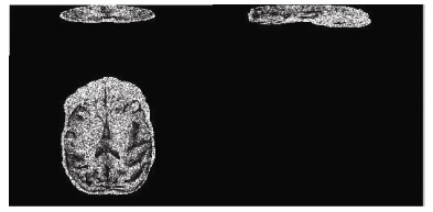
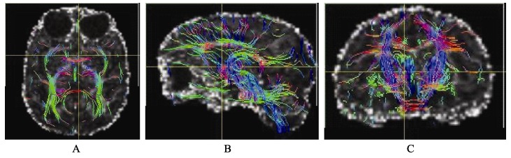
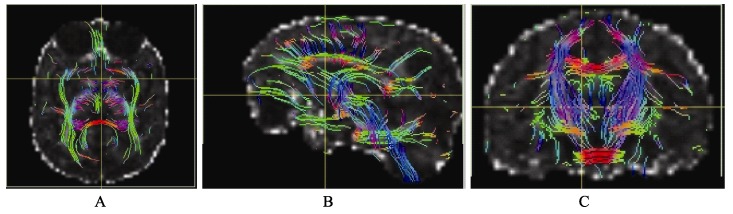
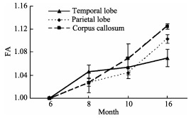
In this study, we analyzed diffusion tensor imaging (DTI) results of brain white matter in rhesus macaques (Macaca mulatta) with four different parameter settings and found that the sequence A (b=1 000 s/mm(2), spatial resolution=1.25 mm×1.25 mm× 1.25 mm, numbers of direction=33, NSA=3) and B (b=800 s/mm(2), spatial resolution=1.25 mm×1.25 mm×1.25 mm, numbers of direction=33, NSA=3) could accurately track coarse fibers. The fractional anisotropy (FA) derived from sequence C (b=1 000s/mm(2), spatial resolution=0.55 mm×0.55 mm×2.5 mm, direction number=33, NSA=3) was too fuzzy to be used in tracking white matter fibers. By comparison, the high resolution and the FA with high contrast of gray matter and white matter derived from sequence D (b=800 s/mm(2), spatial resolution=1.0 mm×1.0 mm ×1.0 mm, numbers of direction=33, NSA=3) qualified in its application in tracking both thick and thin fibers, making it an optimal DTI setting for rhesus macaques.
Keywords: DTI; Rhesus macaque; Whiter matter.
Figures
Similar articles
-
Population-averaged macaque brain atlas with high-resolution ex vivo DTI integrated into in vivo space.Brain Struct Funct. 2017 Dec;222(9):4131-4147. doi: 10.1007/s00429-017-1463-6. Epub 2017 Jun 20. Brain Struct Funct. 2017. PMID: 28634624 Free PMC article.
-
A diffusion-tensor-based white matter atlas for rhesus macaques.PLoS One. 2014 Sep 9;9(9):e107398. doi: 10.1371/journal.pone.0107398. eCollection 2014. PLoS One. 2014. PMID: 25203614 Free PMC article.
-
Artifact correction in diffusion MRI of non-human primate brains on a clinical 3T scanner.J Med Primatol. 2016 Feb;45(1):21-7. doi: 10.1111/jmp.12204. Epub 2015 Dec 22. J Med Primatol. 2016. PMID: 26689605 Free PMC article.
-
The role of diffusion tensor imaging and fractional anisotropy in the evaluation of patients with idiopathic normal pressure hydrocephalus: a literature review.Neurosurg Focus. 2016 Sep;41(3):E12. doi: 10.3171/2016.6.FOCUS16192. Neurosurg Focus. 2016. PMID: 27581308 Review.
-
Diffusion tensor imaging: a review for pediatric researchers and clinicians.J Dev Behav Pediatr. 2010 May;31(4):346-56. doi: 10.1097/DBP.0b013e3181dcaa8b. J Dev Behav Pediatr. 2010. PMID: 20453582 Free PMC article. Review.
Cited by
-
Microstructure changes in whiter matter relate to cognitive impairment in Wilson's disease.Biosci Rep. 2019 Mar 15;39(3):BSR20181651. doi: 10.1042/BSR20181651. Print 2019 Mar 29. Biosci Rep. 2019. PMID: 30804230 Free PMC article.
References
-
- Alexander AL, Hasan KM, Lazar M, Tsuruda JS, Parker DL. 2001. Analysis of partial volume effects in diffusion-tensor MRI. Magnetic Resonance in Medicine, 45 (5): 770- 780. - PubMed
-
- Behrens TEJ, Johansen-Berg H, Woolrich MW, Smith SM, Wheeler-Kingshott CAM, Boulby PA, Barker GJ, Sillery EL, Sheehan K, Ciccarelli O, Thompson AJ, Brady JM, Matthews PM. 2003. Non-invasive mapping of connections between human thalamus and cortex using diffusion imaging. Nature Neuroscience, 6 (7): 750- 757. - PubMed
-
- Jones DK. 2004. The effect of gradient sampling schemes on measures derived from diffusion tensor MRI: a Monte Carlo study. Magnetic Resonance in Medicine, 51 (4): 807- 815. - PubMed
-
- Kim M, Ronen I, Ugurbil K, Kim DS. 2006. Spatial resolution dependence of DTI tractography in human occipito-callosal region. Neuroimage, 32 (3): 1243- 1249. - PubMed
Publication types
MeSH terms
LinkOut - more resources
Full Text Sources
