

Losing the left side of the world: rightward shift in human spatial attention with sleep onset
- PMID: 24867667
- PMCID: PMC4035582
- DOI: 10.1038/srep05092
Losing the left side of the world: rightward shift in human spatial attention with sleep onset
Abstract
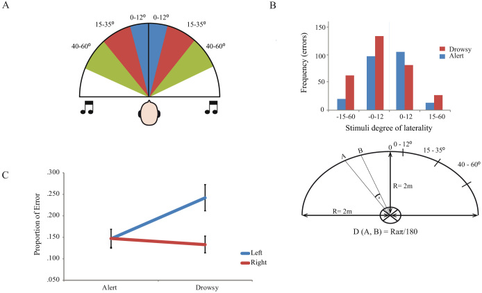
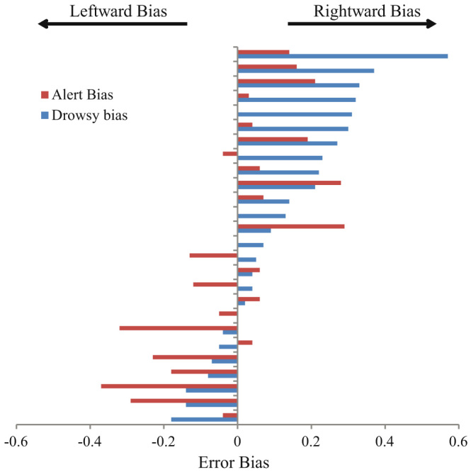
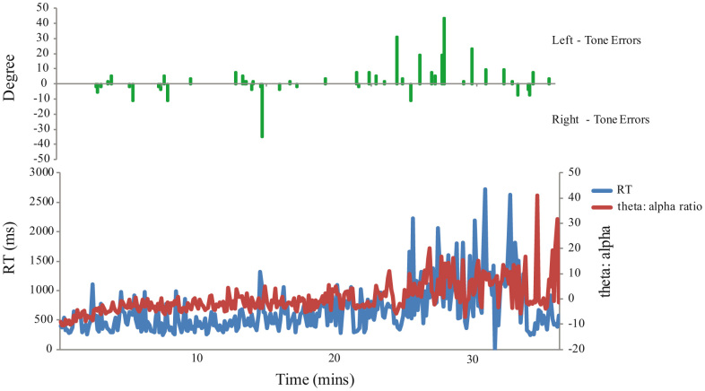
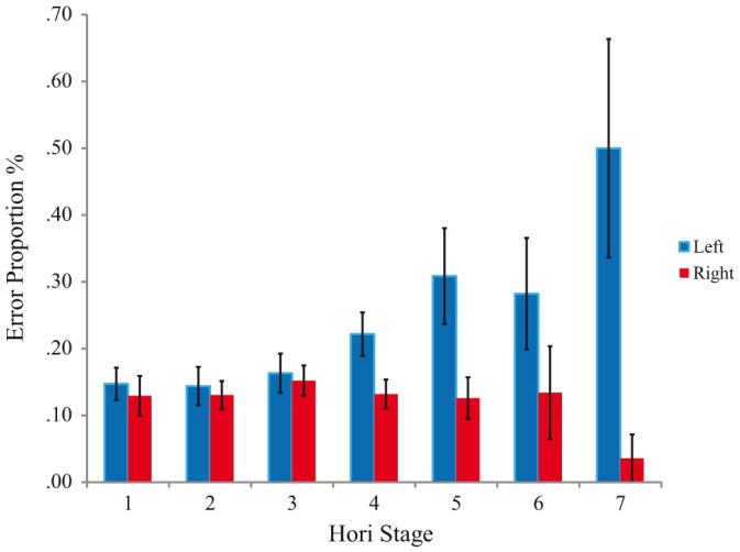
Unilateral brain damage can lead to a striking deficit in awareness of stimuli on one side of space called Spatial Neglect. Patient studies show that neglect of the left is markedly more persistent than of the right and that its severity increases under states of low alertness. There have been suggestions that this alertness-spatial awareness link may be detectable in the general population. Here, healthy human volunteers performed an auditory spatial localisation task whilst transitioning in and out of sleep. We show, using independent electroencephalographic measures, that normal drowsiness is linked with a remarkable unidirectional tendency to mislocate left-sided stimuli to the right. The effect may form a useful healthy model of neglect and help in understanding why leftward inattention is disproportionately persistent after brain injury. The results also cast light on marked changes in conscious experience before full sleep onset.
Figures
Similar articles
-
Does left-handedness confer resistance to spatial bias?Sci Rep. 2015 Mar 17;5:9162. doi: 10.1038/srep09162. Sci Rep. 2015. PMID: 25781078 Free PMC article.
-
Rightward shift in spatial awareness with declining alertness.Neuropsychologia. 2005;43(12):1721-8. doi: 10.1016/j.neuropsychologia.2005.02.009. Epub 2005 Mar 24. Neuropsychologia. 2005. PMID: 16154447
-
Horizontal visual motion modulates focal attention in left unilateral spatial neglect.J Neurol Neurosurg Psychiatry. 1994 Oct;57(10):1228-35. doi: 10.1136/jnnp.57.10.1228. J Neurol Neurosurg Psychiatry. 1994. PMID: 7931385 Free PMC article.
-
Interactions between spatial attention and alertness in healthy adults: A meta-analysis.Cortex. 2019 Oct;119:61-73. doi: 10.1016/j.cortex.2019.03.016. Epub 2019 Mar 30. Cortex. 2019. PMID: 31075552 Review.
-
Different patterns of dissociation in unilateral spatial neglect.Brain Cogn. 1991 Mar;15(2):139-59. doi: 10.1016/0278-2626(91)90023-2. Brain Cogn. 1991. PMID: 2043361 Review.
Cited by
-
A randomized control trial of the effects of home-based online attention training and working memory training on cognition and everyday function in a community stroke sample.Neuropsychol Rehabil. 2022 Dec;32(10):2603-2627. doi: 10.1080/09602011.2021.1972817. Epub 2021 Sep 10. Neuropsychol Rehabil. 2022. PMID: 34505555 Free PMC article. Clinical Trial.
-
Urbanization increases left-bias in line-bisection: an expression of elevated levels of intrinsic alertness?Front Psychol. 2014 Oct 9;5:1127. doi: 10.3389/fpsyg.2014.01127. eCollection 2014. Front Psychol. 2014. PMID: 25346707 Free PMC article.
-
Ocular exposure to blue-enriched light has an asymmetric influence on neural activity and spatial attention.Sci Rep. 2016 Jun 13;6:27754. doi: 10.1038/srep27754. Sci Rep. 2016. PMID: 27291291 Free PMC article.
-
Transient Topographical Dynamics of the Electroencephalogram Predict Brain Connectivity and Behavioural Responsiveness During Drowsiness.Brain Topogr. 2019 Mar;32(2):315-331. doi: 10.1007/s10548-018-0689-9. Epub 2018 Nov 29. Brain Topogr. 2019. PMID: 30498872 Free PMC article.
-
Decreasing Alertness Modulates Perceptual Decision-Making.J Neurosci. 2022 Jan 19;42(3):454-473. doi: 10.1523/JNEUROSCI.0182-21.2021. Epub 2021 Nov 23. J Neurosci. 2022. PMID: 34815316 Free PMC article.
References
-
- Goupil L. & Bekinschtein T. A. Cognitive processing during the transition to sleep. Arch. Ital. Biol. 150, 140–154 (2011). - PubMed
-
- Ogilvie R. D. The process of falling asleep. Sleep Med. Rev. 5, 247–270 (2001). - PubMed
-
- Samann P. G. et al. Development of the brain's default mode network from wakefulness to slow wave sleep. Cereb. Cortex 21, 2082–2093 (2011). - PubMed
-
- Laufs H. et al. Where the BOLD signal goes when alpha EEG leaves. Neuroimage 31, 1408–1418 (2006). - PubMed
Publication types
MeSH terms
Grants and funding
LinkOut - more resources
Full Text Sources
Other Literature Sources
Miscellaneous
