. 2014 May;26(5):2249-2264.
doi: 10.1105/tpc.114.123737. Epub 2014 May 27.

OPT3 Is a Phloem-Specific Iron Transporter That Is Essential for Systemic Iron Signaling and Redistribution of Iron and Cadmium in Arabidopsis

Affiliations

OPT3 Is a Phloem-Specific Iron Transporter That Is Essential for Systemic Iron Signaling and Redistribution of Iron and Cadmium in Arabidopsis

Zhiyang Zhai et al. Plant Cell. 2014 May.

Abstract

Iron is essential for both plant growth and human health and nutrition. Knowledge of the signaling mechanisms that communicate iron demand from shoots to roots to regulate iron uptake as well as the transport systems mediating iron partitioning into edible plant tissues is critical for the development of crop biofortification strategies. Here, we report that OPT3, previously classified as an oligopeptide transporter, is a plasma membrane transporter capable of transporting transition ions in vitro. Studies in Arabidopsis thaliana show that OPT3 loads iron into the phloem, facilitates iron recirculation from the xylem to the phloem, and regulates both shoot-to-root iron signaling and iron redistribution from mature to developing tissues. We also uncovered an aspect of crosstalk between iron homeostasis and cadmium partitioning that is mediated by OPT3. Together, these discoveries provide promising avenues for targeted strategies directed at increasing iron while decreasing cadmium density in the edible portions of crops and improving agricultural productivity in iron deficient soils.


Figures

Figure 1.
Figure 1.
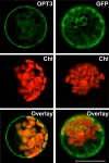
OPT3 Localizes to the Plasma Membrane in Arabidopsis Protoplasts. GFP-mediated fluorescence, derived from the OPT3-GFP (OPT3) constructor or the empty GFP vector (GFP), and chlorophyll autofluorescence (Chl) were visualized using FITC or rhodamine filter sets. Superimposed images of chlorophyll autofluorescence and GFP-mediated fluorescence (Overlay) were created to demonstrate that green fluorescence was derived from GFP. Bar = 20 μm.
Figure 2.
Figure 2.
OPT3 Does Not Mediate GSH Transport in S. cerevisiae. (A) S. cerevisiae opt1 mutant cells expressing Sc-OPT1cDNA (open circles), the empty vector (closed circles), or vector with the At-OPT3 cDNA insert (open squares) were grown in SC-S media supplemented without or with 200 μM GSH. OD values were measured after 24 and 48 h of culturing at 30°C. (B) Time course of in vitro [35S]GSH uptake by opt1 mutant cells expressing Sc-OPT1cDNA (open circles), the empty vector (closed circles), or vector with the At-OPT3 cDNA insert (open squares). Error bars represent se (n = 3).
Figure 3.
Figure 3.
OPT3 Partially Rescues Fe Deficiency of the fet3 fet4 S. cerevisiae Mutant. (A) The wild type and the fet3 fet4 mutant, transformed with the empty vector (Wt and fet3fet4, respectively), and the fet3 fet4 mutant transformed with At-OPT3 or At-IRT1 cDNAs (OPT3 and IRT1) were serially 10-fold diluted and spotted onto solid medium supplemented with the indicated concentrations of the Fe chelator BPS. Colonies were visualized after incubating plates for 6 d at 30°C. Dilution series are indicated on the left. (B) Iron concentration in different S. cerevisiae lines, designated as in (A). Shown are mean values ± se (n = 5 to 8); asterisks indicate statistically significant differences (P ≤ 0.001) from the empty vector–expressing opt2 or fet3 fet4 mutant cells.
Figure 4.
Figure 4.
OPT3 Is Functional in X. laevis Oocytes. (A) Resting membrane potentials of OPT3-expressing (OPT3) and water-injected (Control) cells measured in standard ND96 recording solution. (B) Example of OPT3-mediated currents (right panel) elicited in response to holding potentials ranging from 0 to −140 mV (shown only in 20-mV increments for clarity) in standard ND96 recording solution. Endogenous currents recorded in control cells are shown for reference on the left panel. The arrow and dotted line on the left margin indicates the zero current level. (C) Mean current-voltage (I/V) curves constructed from the steady state currents recordings such as those shown in (B) for holding potentials ranging from 0 to −140 mV in 10-mV steps. Error bars represent se (n = 5).
Figure 5.
Figure 5.
OPT3 Mediates Cation Uptake in X. laevis Oocytes. Uptake of Fe2+ (A) and Cd2+ (B) in OPT3-expressing oocytes (OPT3) or water-injected cells (Control) at different time points. The basal uptake solution supplemented with 0.4 mM FeSO4 + 1 mM l-ascorbic acid (A) or 0.5 mM CdCl2 (B), yielded free extracellular ionic activities of 35 and 150 μM of {Cd2+}out and {Fe2+}out, respectively, as determined by GEOCHEM-EZ (Shaff et al., 2010). The dashed line in (A) indicates the basal concentration of endogenous Fe2+. Error bars represent se (n = 5). Asterisks indicate statistically significant differences (P ≤ 0.05).
Figure 6.
Figure 6.
Tissue- and Cell-Type Specificity of OPT3 Expression in Arabidopsis. (A) to (E) Histochemical analysis of the OPT3 promoter activity in transgenic plants expressing the OPT3pro-GUS construct. (A) Representative expression pattern for OPT3 in a whole seedling. Note the bulk of OPT3 expression in shoots (main figure) but not in roots (inset). (B) Close-up of the leaf area to demonstrate OPT3 expression in minor veins. (C) Expression of OPT3 at the node (arrow). (D) and (E) Pattern of OPT3 expression in reproductive organs. (F) to (M) Hand-cut cross sections through the petiole ([F] to [I]) or inflorescence stem ([J] to [M]) of 21-d-old transgenic plants expressing the OPT3pro-GFP construct. (F) and (J) Differential interference contrast images of sections through the petiole and stem at the nodal region, respectively. Overlay images ([I] and [M]) were created to show that GFP-mediated fluorescence ([G] and [K]) does not overlap with phenolics-mediated autofluorescence in xylem vessels and chlorophyll-mediated autofluorescence in parenchyma cells ([H] and [L]). Xy, xylem; Ph, phloem. Bar = 100 μm.
Figure 7.
Figure 7.
OPT3 Mediates Fe Transport from Source to Sink Tissues. ICP-MS analysis of Fe and Cd concentrations in old and young leaves ([A] to [C]) and seeds (D) of wild-type and opt3-3 mutant plants. Asterisks indicate statistically significant differences from the corresponding leaves in the wild type (P ≤ 0.001). Letters (a and aa) indicate statistically significant differences between old and young rosette leaves of the opt3-3 mutant (aP ≤ 0.05 or aaP ≤ 0.01). Statistically significant differences between old and young rosette leaves in the wild type are indicated as (bbP ≤ 0.001). DW, dry weight. (A) to (C) Young and old rosette leaves were harvested at the late vegetative stage from hydroponically grown plants. In (A), plants were grown in 10 μM 56FeHBED (Fe) until tissues were collected for the ICP-MS analysis. In (B), plants were grown in 56Fe until the late vegetative stage and then transferred for 24 h to a fresh hydroponic medium containing 25 μM 57FeHBED (57Fe), while in (C), plants were grown for additional 24 h with 25 μM CdCl2 before sink and source leaves were harvested and subjected to the ICP-MS analysis. (D) Iron and Cd concentrations in plants grown in soil with 7.5 nM Cd or 10 μM Fe. Error bars represent se (n = 3).
Figure 8.
Figure 8.
OPT3 Mediates Xylem-to-Phloem Fe Transfer. (A) and (B) The concentration of Fe and K in phloem sap (A) or Fe and Cd in xylem sap (B), both collected from wild-type and opt3-3 mutant plants grown hydroponically under Fe-sufficient conditions. Error bars represent se (n = 3). Asterisks indicate statistically significant differences (*P ≤ 0.05 and **P ≤ 0.001). (C) Synchrotron x-ray fluorescence map of Fe distribution in leaves of the wild type and the opt3-3 mutant in Arabidopsis. Note that the line of Fe in the leaf of the wild type is an artifact of the leaf folding. Bar = 1 mm.
Figure 9.
Figure 9.
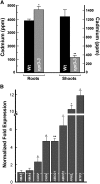
Leaves of opt3-3 Plants Accumulate Less Cd Than Leaves of Wild-Type Plants. (A) The concentration of Cd in roots and shoots of wild-type and opt3-3 mutant plants grown for 4 d with 25 μM CdCl2. Shown are mean values ± se; n = 3. Asterisks indicate statistically significant differences (*P ≤ 0.05 and **P ≤ 0.001). (B) Transcript abundance of the indicated genes was analyzed in roots of wild-type and opt3-3 plants, both grown hydroponically to the vegetative stage. Results are presented relative to expression of these genes in roots of wild-type plants designated as 1 (dashed line). Error bars indicate se (n = 6). Asterisks indicate statistically significant differences (*P ≤ 0.05 and **P ≤ 0.001, respectively).
Figure 10.
Figure 10.
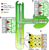
Model of OPT3 Function in Arabidopsis. This figure summarizes the proposed dual roles of OPT3 in the redistribution of Fe from source to sink tissues, in shoot-to-root signaling of shoot Fe status and its contribution to Cd partitioning in Arabidopsis. (A) OPT3 is expressed in CCs of the phloem that might differentiate into the transfer cells (TF) in minor veins of leaves and in nodes of stems. In these sites, OPT3 facilitates Fe loading into the phloem, possibly by direct xylem-to-phloem Fe transport. (B) At the whole-plant level, OPT3 is involved in redistribution of Fe from sources to sinks and Fe recirculation into roots. Iron recirculation into roots via OPT3 plays a signaling role by conveying Fe status of the shoot. (C) Although OPT3 transports Cd in vitro, it is likely that it mediates root-to-shoot partitioning of Cd indirectly by orchestrating shoot-to-root Fe signaling that, in turn, alters expression of genes encoding multispecific transition ion transporters (e.g., FPN2, MTP3, CAX2/4, ABCC1/2, and possibly others). These transporters facilitate vacuolar sequestration of Cd and its retention in the root, which in turn, affects root-to-shoot Cd partitioning and Cd resistance. XP, xylem parenchyma cells.

Similar articles

Cited by

References

    1. Alonso J.M., et al. (2003). Genome-wide insertional mutagenesis of Arabidopsis thaliana. Science 301: 653–657 - PubMed
    1. Amasheh S., Weber W. (1999). Further characteristics of the Ca(2+)-inactivated Cl(-) channel in Xenopus laevis oocytes. J. Membr. Biol. 172: 169–179 - PubMed
    1. Andriunas F.A., Zhang H.-M., Xia X., Patrick J.W., Offler C.E. (2013). Intersection of transfer cells with phloem biology-broad evolutionary trends, function, and induction. Front Plant Sci 4: 221. - PMC - PubMed
    1. Arrivault S., Senger T., Krämer U. (2006). The Arabidopsis metal tolerance protein AtMTP3 maintains metal homeostasis by mediating Zn exclusion from the shoot under Fe deficiency and Zn oversupply. Plant J. 46: 861–879 - PubMed
    1. Arteca R.N., Arteca J.M. (2000). A novel method for growing Arabidopsis thaliana plants hydroponically. Physiol. Plant. 108: 188–193

LinkOut - more resources