. 2014 Jul 11;289(28):19435-47.
doi: 10.1074/jbc.M114.553867. Epub 2014 May 27.

Directed expression of a chimeric type II keratin partially rescues keratin 5-null mice

Affiliations

Directed expression of a chimeric type II keratin partially rescues keratin 5-null mice

David M Alvarado et al. J Biol Chem. .

Abstract

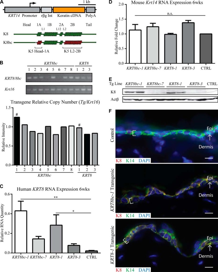
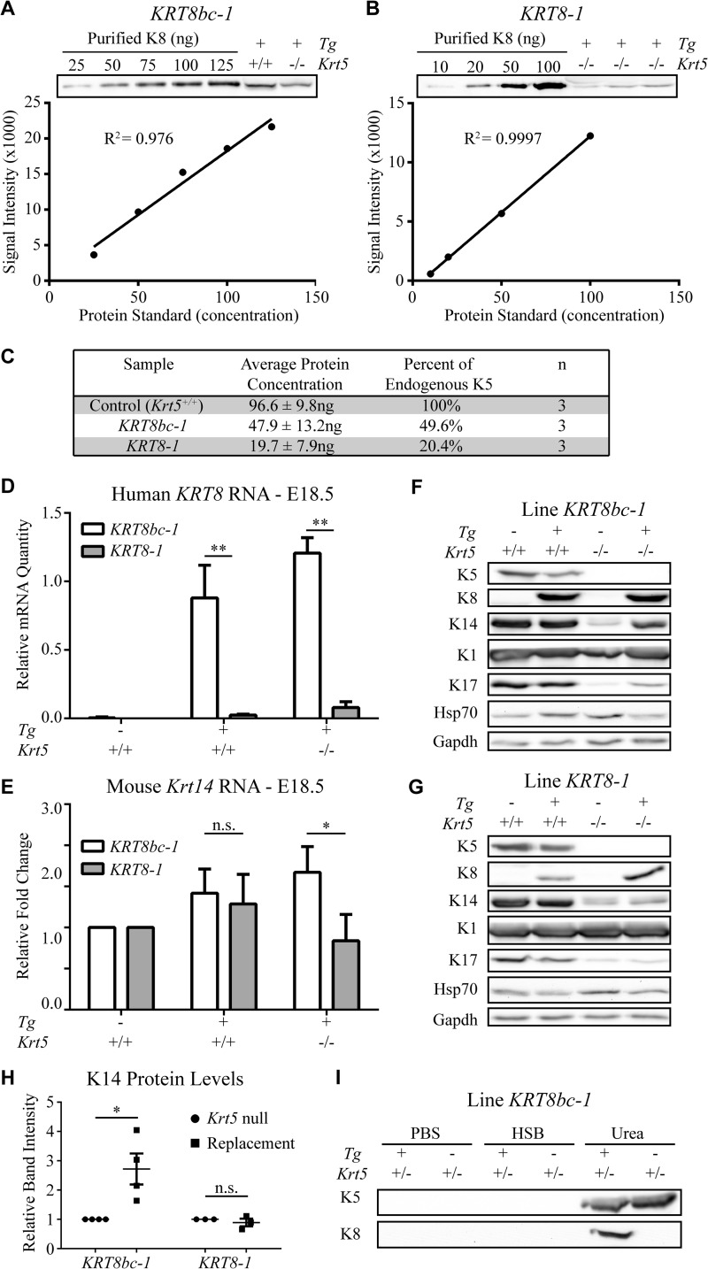
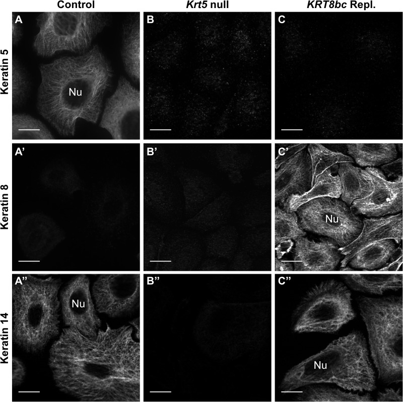
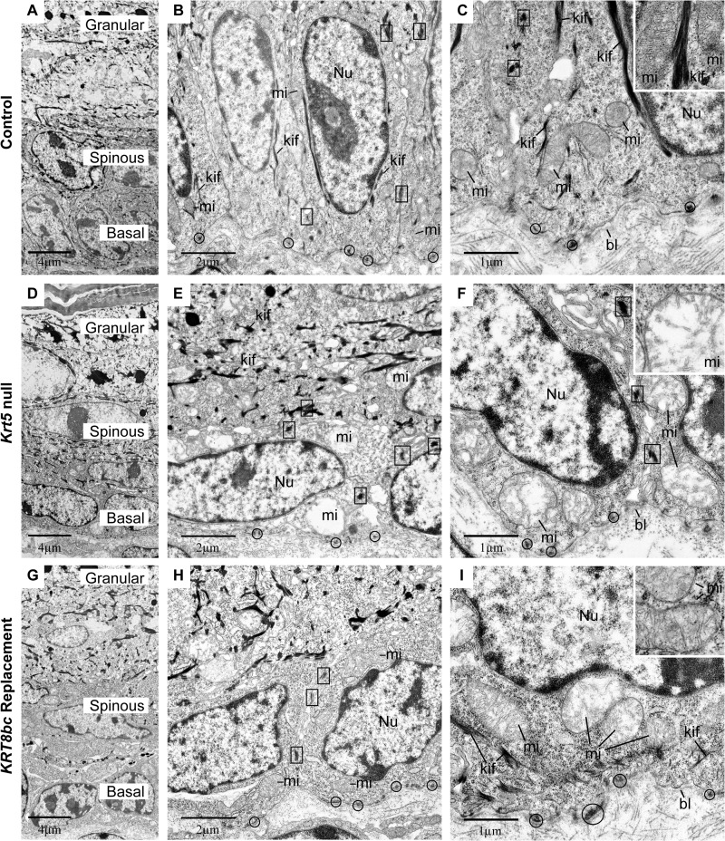
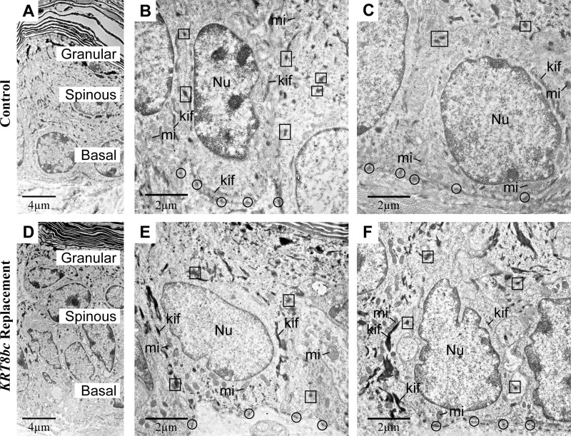
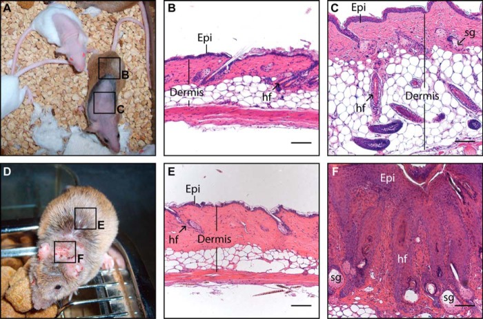
The crucial role of structural support fulfilled by keratin intermediate filaments (IFs) in surface epithelia likely requires that they be organized into cross-linked networks. For IFs comprised of keratins 5 and 14 (K5 and K14), which occur in basal keratinocytes of the epidermis, formation of cross-linked bundles is, in part, self-driven through cis-acting determinants. Here, we targeted the expression of a bundling-competent KRT5/KRT8 chimeric cDNA (KRT8bc) or bundling-deficient wild type KRT8 as a control to the epidermal basal layer of Krt5-null mice to assess the functional importance of keratin IF self-organization in vivo. Such targeted expression of K8bc rescued Krt5-null mice with a 47% frequency, whereas K8 completely failed to do so. This outcome correlated with lower than expected levels of K8bc and especially K8 mRNA and protein in the epidermis of E18.5 replacement embryos. Ex vivo culture of embryonic skin keratinocytes confirmed the ability of K8bc to form IFs in the absence of K5. Additionally, electron microscopy analysis of E18.5 embryonic skin revealed that the striking defects observed in keratin IF bundling, cytoarchitecture, and mitochondria are partially restored by K8bc expression. As young adults, viable KRT8bc replacement mice develop alopecia and chronic skin lesions, indicating that the skin epithelia are not completely normal. These findings are consistent with a contribution of self-mediated organization of keratin IFs to structural support and cytoarchitecture in basal layer keratinocytes of the epidermis and underscore the importance of context-dependent regulation for keratin genes and proteins in vivo.

Keywords: Blistering; Cell Fragility; Epidermis; Epidermolysis Bullosa Simplex; Genodermatosis; Intermediate Filament; Keratin; Mitochondria; Skin; Transgenic Mice.


Figures

FIGURE 1.
FIGURE 1.
Generation and characterization of transgenes. A, schematic depiction of the transgenes used in this study. The human KRT14 gene promoter was used to drive the tissue-specific expression of keratin cDNAs (KRT8 and KRT8bc), and the rabbit β-globin intron (rβg Int) and human KRT14 3′-UTR sequence (Poly(A)) serve to stabilize the transgene mRNA in mouse cells. B, relative transgene copy number analysis was performed by conventional PCR of genomic DNA with transgene-specific primers. Krt16 was used as a single copy number reference gene. # indicates the highest relative copy number line for each transgene. C and D, quantitative RT-PCR analysis of transgenic human KRT8 mRNA (C) and endogenous mouse Krt14 mRNA (D) in back skin harvested from sex-matched 6-week-old animals. Relative RNA amount is normalized to both actin and Gapdh. CTRL indicates Krt5+/+ skin. Error bars represent S.E. A one-way analysis of variance (Dunnett's test) was used to test for significance, and the adjusted p values are reported. n.s., not significant; *, p < 0.04; **, p < 0.002. E, analysis of total skin protein extracts (10 μg/lane) by Western blotting in 6-week-old sex-matched adult animals. Two mice were analyzed for each of four transgenic lines; CTRL indicates Krt5+/+ skin. F, analysis of transgene expression in frozen skin sections of 6-week-old sex-matched adult animals. K8 epitopes are only present in basal layer keratinocytes (see brackets) of KRT8bc-1 and KRT8-1 transgenic epidermis and co-localizes with endogenous K14 in a normal keratin filament network. Control indicates Krt5+/+ skin. Arrows depict the interface between the epidermis (Epi) and dermis. Bars, 10 μm. wks, weeks.
FIGURE 2.
FIGURE 2.
Analysis of transgene expression in E18.5 mouse embryos. A and B, quantification of transgenic protein expressed in KRT8bc line 1 (KRT8bc-1; A) and KRT8 line 1 (KRT8-1; B) using standard curves of purified recombinant human K8. The line of best fit and R2 values were calculated using GraphPad Prism. C, summary of the transgenic protein quantification from multiple biological (n) and technical replicates. D, transgene RNA expression levels for lines KRT8bc-1 and KRT8-1 in E18.5 skin. E, endogenous Krt14 RNA expression levels for lines KRT8bc-1 and KRT8-1 in E18.5 skin. RNA levels are illustrated relative to actin and Gapdh. Error bars represent S.E. A two-way analysis of variance (Sidak's test) was used to test for significance, and the adjusted p values are reported. n.s., not significant; *, p < 0.005; **, p < 0.0001. F and G, expression of transgenic and endogenous proteins for lines KRT8bc-1 (F) and KRT8-1 (G) in E18.5 skin is shown by Western blot analysis. H, intensity of K14 protein signal relative to loading control signal was quantified, and the increase of K14 in the replacement mouse skin setting is illustrated. I, Western blot analysis of high salt extraction performed on KRT8bc-1 E18.5 skin. Even in the presence of endogenous K5 as a competitor, transgenic K8bc was still found in the insoluble pool. HSB, high salt buffer.
FIGURE 3.
FIGURE 3.
Analysis of embryonic skin keratinocytes in primary culture by indirect immunofluorescence. Conventional fluorescence microscopy of keratinocytes harvested from Krt5+/+ (Control; A–A″), Krt5−/− (Krt5 null; B–B″), or KRT8bcTg/−Krt5−/− (KRT8bc Repl.; C–C″) E18.5 embryos (as indicated on top) is shown. Fixed cells were immunostained for keratin 5, keratin 8, and keratin 14 epitopes as indicated at left. Nu, nucleus. Bars, 10 μm.
FIGURE 4.
FIGURE 4.
Ultrastructural analysis of E18.5 epidermis in situ. Krt5+/+ (Control; A–C), Krt5−/− (Krt5 null; D–F), and KRT8bcTg/−Krt5−/− (KRT8bc Replacement; G–I) mouse epidermis at E18.5 was analyzed by routine transmission electron microscopy of thin sections. A, D, and G provide low magnification surveys of the living layers of epidermis (basal, spinous, and granular), whereas all other panels provide details of basal keratinocytes. bl, basal lamina; kif, keratin intermediate filament bundles; mi, mitochondria; Nu, nucleus. Examples of hemidesmosomes and desmosomes are circled and boxed, respectively, and representative mitochondria are detailed in the insets. Bars, 4 μm in A, D, and G; 2 μm in B, E, and H; and 1 μm in C, F, and I.
FIGURE 5.
FIGURE 5.
Ultrastructural analysis of adult control and KRT8bc replacement epidermis in situ. Krt5+/+ (Control; A–C), and KRT8bcTg/−Krt5−/− (KRT8bc Replacement; D–F) epidermis in 11-week-old mice was analyzed by routine transmission electron microscopy of thin sections. A and D provide low magnification surveys of the living layers of epidermis (basal, spinous, and granular), whereas B, C, E, and F provide details of basal keratinocytes. kif, keratin intermediate filament bundles; mi, mitochondria; Nu, nucleus. Examples of hemidesmosomes and desmosomes are circled and boxed, respectively. Bars, 4 μm in A and D and 2 μm in B, C, E, and F.
FIGURE 6.
FIGURE 6.
Analysis of late onset phenotypes in adult KRT8bc replacement animals. A macroscopic survey (A and D) and histology (B, C, E, and F) illustrating the late onset phenotypes arising in KRT8bc replacement animals are shown. A, 6-month-old adult KRT8bc replacement animals displaying both normal skin and areas of alopecia are shown. B, hairy skin from KRT8bc replacement animal (see boxB” in A) showing normal histology. C, phenotypic skin sample from KRT8bc replacement animal (see boxC” in A) showing misoriented, anagen-staged hair follicles and thickened epidermis. D, a 3-month-old adult KRT8bc replacement animal showing both normal skin and areas of inflamed and hyperkeratotic, scaly skin. E, hairy skin from KRT8bc replacement animal showing normal histology (see boxE” in D). F, phenotypic skin from replacement animals (see boxF” in D) depicting hyperplastic epidermis, pilosebaceous cysts, and a high level of dermal infiltration, suggesting an inflammatory and immune infiltration. Epi, epidermis; hf, hair follicle; sg, sebaceous gland. Bars, 100 μm.

References

    1. Omary M. B., Coulombe P. A., McLean W. H. (2004) Intermediate filament proteins and their associated diseases. N. Engl. J. Med. 351, 2087–2100 - PubMed
    1. Szeverenyi I., Cassidy A. J., Chung C. W., Lee B. T., Common J. E., Ogg S. C., Chen H., Sim S. Y., Goh W. L., Ng K. W., Simpson J. A., Chee L. L., Eng G. H., Li B., Lunny D. P., Chuon D., Venkatesh A., Khoo K. H., McLean W. H., Lim Y. P., Lane E. B. (2008) The Human Intermediate Filament Database: comprehensive information on a gene family involved in many human diseases. Hum. Mutat. 29, 351–360 - PubMed
    1. Coulombe P. A., Kerns M. L., Fuchs E. (2009) Epidermolysis bullosa simplex: a paradigm for disorders of tissue fragility. J. Clin. Investig. 119, 1784–1793 - PMC - PubMed
    1. Bonifas J. M., Rothman A. L., Epstein E. H., Jr. (1991) Epidermolysis bullosa simplex: evidence in two families for keratin gene abnormalities. Science 254, 1202–1205 - PubMed
    1. Coulombe P. A., Hutton M. E., Letai A., Hebert A., Paller A. S., Fuchs E. (1991) Point mutations in human keratin 14 genes of epidermolysis bullosa simplex patients: genetic and functional analyses. Cell 66, 1301–1311 - PubMed

Publication types

MeSH terms

Supplementary concepts

LinkOut - more resources