

Whole-genome analyses of Enterococcus faecium isolates with diverse daptomycin MICs
- PMID: 24867964
- PMCID: PMC4136017
- DOI: 10.1128/AAC.02686-14
Whole-genome analyses of Enterococcus faecium isolates with diverse daptomycin MICs
Abstract
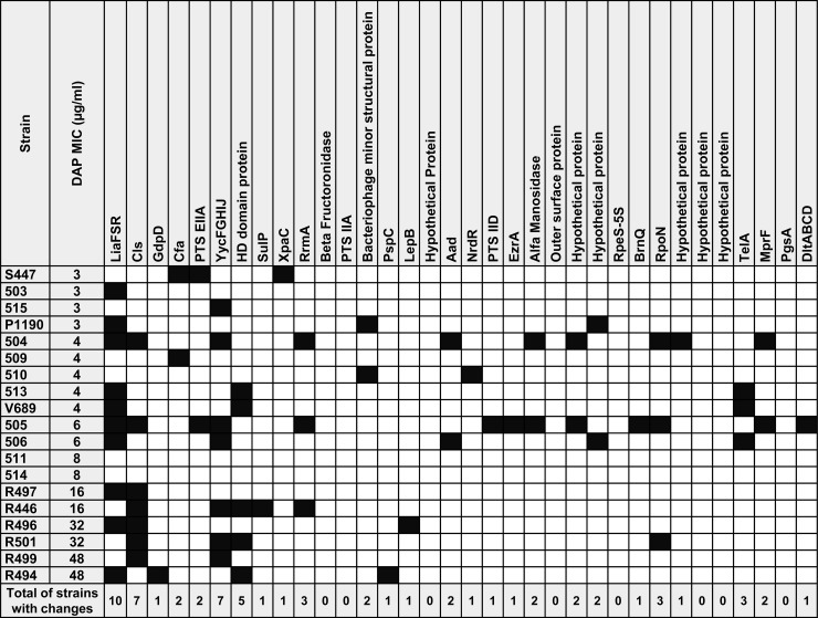
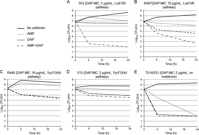
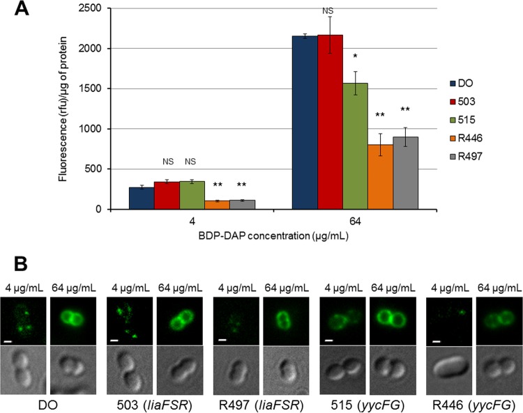
Daptomycin (DAP) is a lipopeptide antibiotic frequently used as a "last-resort" antibiotic against vancomycin-resistant Enterococcus faecium (VRE). However, an important limitation for DAP therapy against VRE is the emergence of resistance during therapy. Mutations in regulatory systems involved in cell envelope homeostasis are postulated to be important mediators of DAP resistance in E. faecium. Thus, in order to gain insights into the genetic bases of DAP resistance in E. faecium, we investigated the presence of changes in 43 predicted proteins previously associated with DAP resistance in enterococci and staphylococci using the genomes of 19 E. faecium with different DAP MICs (range, 3 to 48 μg/ml). Bodipy-DAP (BDP-DAP) binding to the cell membrane assays and time-kill curves (DAP alone and with ampicillin) were performed. Genetic changes involving two major pathways were identified: (i) LiaFSR, a regulatory system associated with the cell envelope stress response, and (ii) YycFGHIJ, a system involved in the regulation of cell wall homeostasis. Thr120 → Ala and Trp73 → Cys substitutions in LiaS and LiaR, respectively, were the most common changes identified. DAP bactericidal activity was abolished in the presence of liaFSR or yycFGHIJ mutations regardless of the DAP MIC and was restored in the presence of ampicillin, but only in representatives of the LiaFSR pathway. Reduced binding of BDP-DAP to the cell surface was the predominant finding correlating with resistance in isolates with DAP MICs above the susceptibility breakpoint. Our findings suggest that genotypic information may be crucial to predict response to DAP plus β-lactam combinations and continue to question the DAP breakpoint of 4 μg/ml.
Copyright © 2014, American Society for Microbiology. All Rights Reserved.
Figures
References
-
- Lebreton F, van Schaik W, McGuire AM, Godfrey P, Griggs A, Mazumdar V, Corander J, Cheng L, Saif S, Young S, Zeng Q, Wortman J, Birren B, Willems RJ, Earl AM, Gilmore MS. 2013. Emergence of epidemic multidrug-resistant Enterococcus faecium from animal and commensal strains. mBio 4:e00534–13. 10.1128/mBio.00534-13 - DOI - PMC - PubMed
-
- Arias CA, Torres HA, Singh KV, Panesso D, Moore J, Wanger A, Murray BE. 2007. Failure of daptomycin monotherapy for endocarditis caused by an Enterococcus faecium strain with vancomycin-resistant and vancomycin-susceptible subpopulations and evidence of in vivo loss of the vanA gene cluster. Clin. Infect. Dis. 45:1343–1346. 10.1086/522656 - DOI - PubMed
Publication types
MeSH terms
Substances
Grants and funding
LinkOut - more resources
Full Text Sources
Other Literature Sources
Medical
