Rare cause of abdominal incidentaloma: Hepatoduodenal ligament teratoma
- PMID: 24868330
- PMCID: PMC4033282
- DOI: 10.4240/wjgs.v6.i5.80
Rare cause of abdominal incidentaloma: Hepatoduodenal ligament teratoma
Abstract
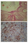
The occurrence of a hepatoduodenal ligament teratoma is extremely rare, with only a few cases reported in the literature. This case report describes the discovery of a hepatoduodenal ligament lesion revealed during abdominal ultrasonography for cholelithiasis-related abdominal pain in a 27-year-old female. Cross-sectional imaging identified a 5 cm × 4 cm heterogeneous mass of fat tissue with irregular calcification located in the posterior-superior aspect of the head of the pancreas. An encapsulated lesion showing no invasion to the common bile duct or adjacent organs and vessels was exposed during laparotomy and resected. Intraoperative cholangiography during the cholecystectomy showed no abnormalities. The postoperative course was uneventful. Pathological analysis of the resected mass indicated hepatoduodenal ligament teratoma. This case report demonstrates that cross-sectional imaging, such as computed tomography, can reveal suspected incidences of this rare type of teratoma, which can then be confirmed after pathologic analysis of the specimen. The prognosis after complete surgical resection of lesions presenting with benign pathological features is excellent.
Keywords: Abdominal incidentaloma; Hepatobiliary surgery; Hepatoduodenal ligament; Surgery; Teratoma.
Figures


References
-
- Engel RM, Elkins RC, Fletcher BD. Retroperitoneal teratoma. Review of the literature and presentation of an unusual case. Cancer. 1968;22:1068–1073. - PubMed
-
- Frexes M, Neblett WW, Holcomb GW. Spectrum of biliary disease in childhood. South Med J. 1986;79:1342–1349. - PubMed
-
- Akimov OV. [Hepatoduodenal ligament teratoma followed by hypertensive syndrome of the portal vein] Arkh Patol. 1989;51:60–62. - PubMed
-
- Kim WS, Choi BI, Lee YS, Chi JG, Park HR, Kim I, Yeon KM, Han MC. Endodermal sinus tumour associated with benign teratoma of the common bile duct. Pediatr Radiol. 1993;23:59–60. - PubMed
-
- Demircan M, Uguralp S, Mutus M, Kutlu R, Mizrak B. Teratoma arising from anomalous common bile ducts: a case report. J Pediatr Surg. 2004;39:e1–e2. - PubMed
Publication types
LinkOut - more resources
Full Text Sources
Other Literature Sources

