A case of juvenile huntington disease in a 6-year-old boy
- PMID: 24868381
- PMCID: PMC4027671
- DOI: 10.14802/jmd.10012
A case of juvenile huntington disease in a 6-year-old boy
Abstract
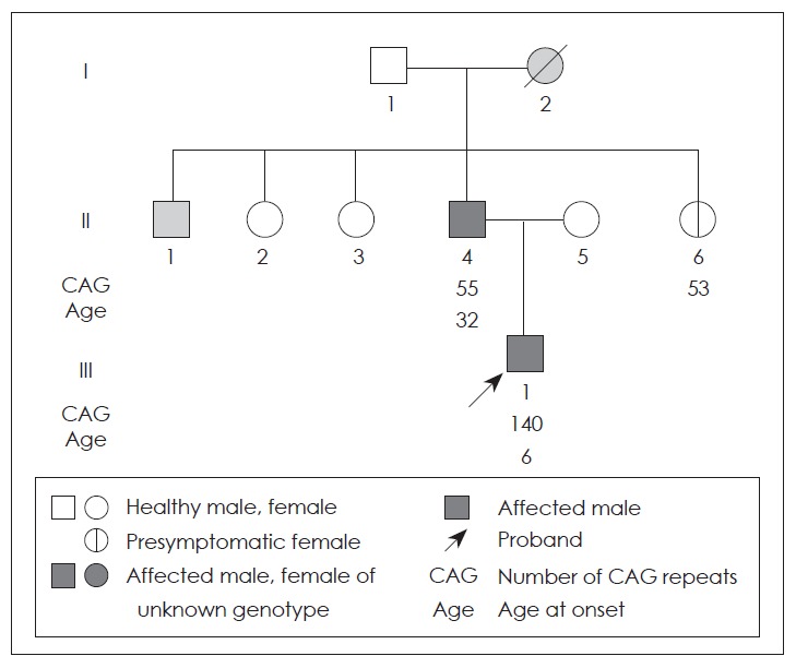
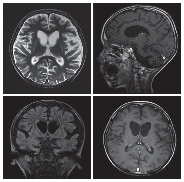
Huntington disease is a neurodegenerative disorder distinguished by the triad of dominant inheritance, choreoathetosis and dementia, usually with onset in the fourth and fifth decades. It is caused by an unstable cytosine-adenine-guanine (CAG) trinucleotide repeat expansion in the gene IT15 in locus 4p16.3. Juvenile HD that constitutes about 3% to 10% of all patients is clinically different from adult-onset form and characterized by a larger number of CAG repeats typically exceeding 60. We report a case of a 6-year-old boy with myoclonic seizure and 140 CAG repeats confirmed by molecular genetic analysis.
Keywords: Juvenile Huntington disease; Seizure; Trinucleotide repeat expansions.
Figures
References
-
- A novel gene containing a trinucleotide repeat that is expanded and unstable on Huntington’s disease chromosomes. The Huntington’s Disease Collaborative Research Group. Cell. 1993;72:971–983. - PubMed
-
- Nance MA, Myers RH. Juvenile onset Huntington’s disease--clinical and research perspectives. Ment Retard Dev Disabil Res Rev. 2001;7:153–157. - PubMed
-
- Lee MS, Shin CH, Son DW, Park KH, Kim DH, Kim KY, et al. A case of juvenile Huntington’s disease confirmed by molecular genetic analysis. J Korean Child Neurol Soc. 1999;7:113–118.
-
- Wojaczyńska-Stanek K, Adamek D, Marszał E, Hoffman-Zacharska D. Huntington disease in a 9-year-old boy: clinical course and neuropathologic examination. J Child Neurol. 2006;21:1068–1073. - PubMed
-
- Nance MA. Genetic testing of children at risk for Huntington’s disease. US Huntington Disease Genetic Testing Group. Neurology. 1997;49:1048–1053. - PubMed
LinkOut - more resources
Full Text Sources