

The osteogenesis of bone marrow stem cells on mPEG-PCL-mPEG/hydroxyapatite composite scaffold via solid freeform fabrication
- PMID: 24868523
- PMCID: PMC4020560
- DOI: 10.1155/2014/321549
The osteogenesis of bone marrow stem cells on mPEG-PCL-mPEG/hydroxyapatite composite scaffold via solid freeform fabrication
Abstract
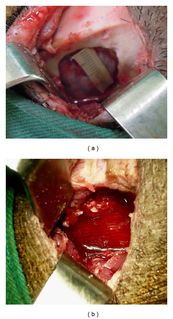
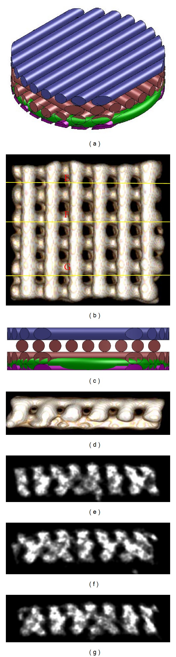
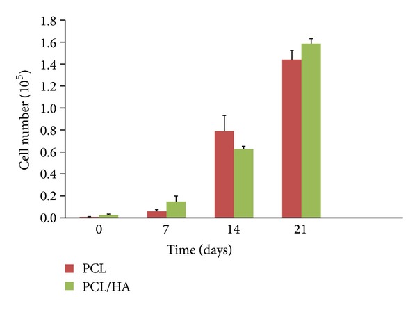
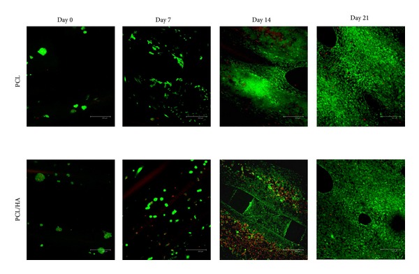
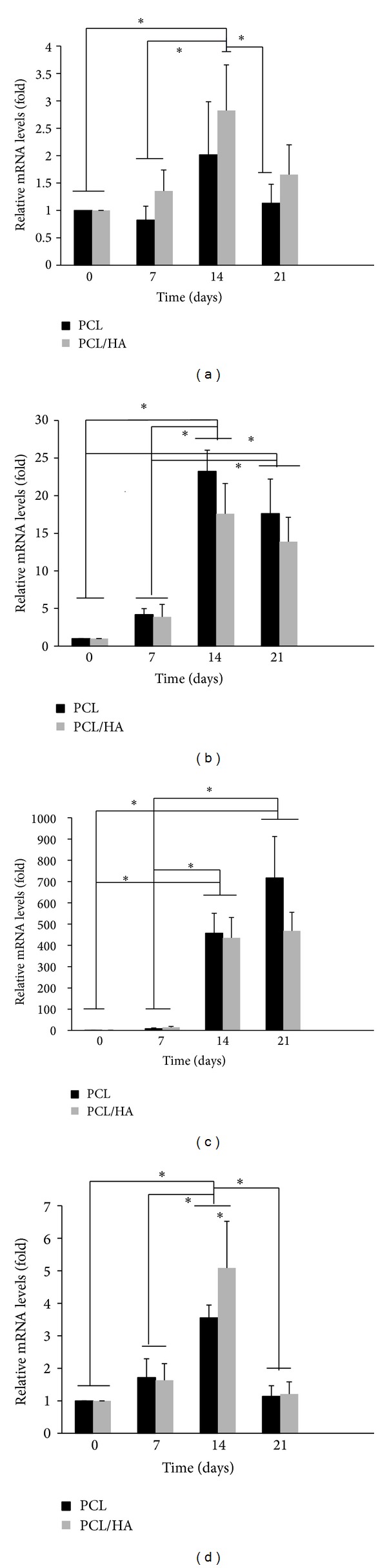
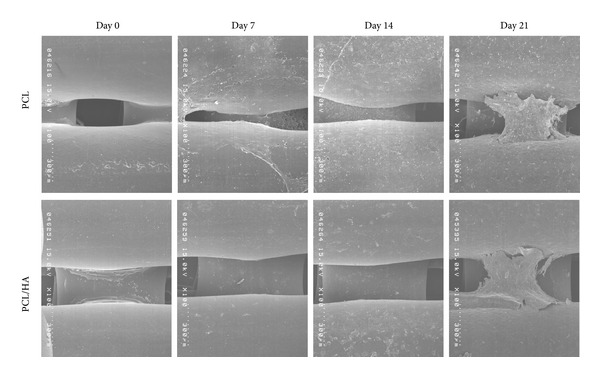
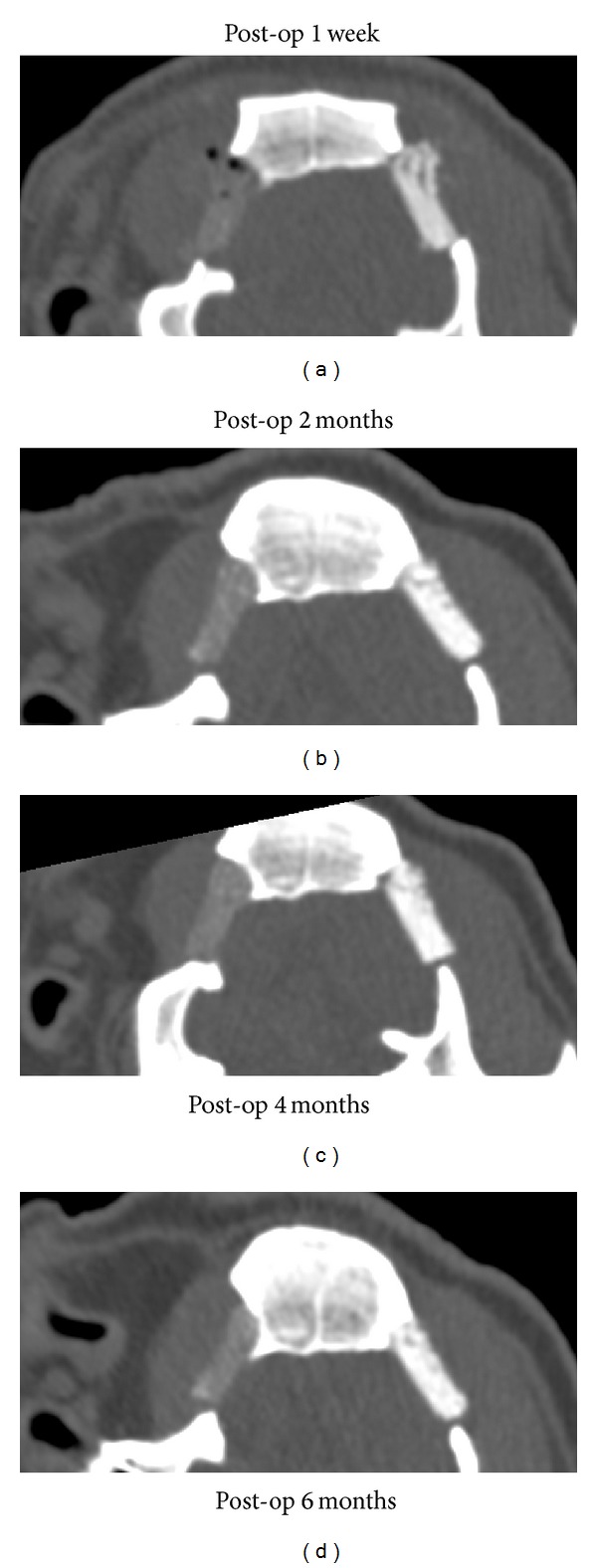
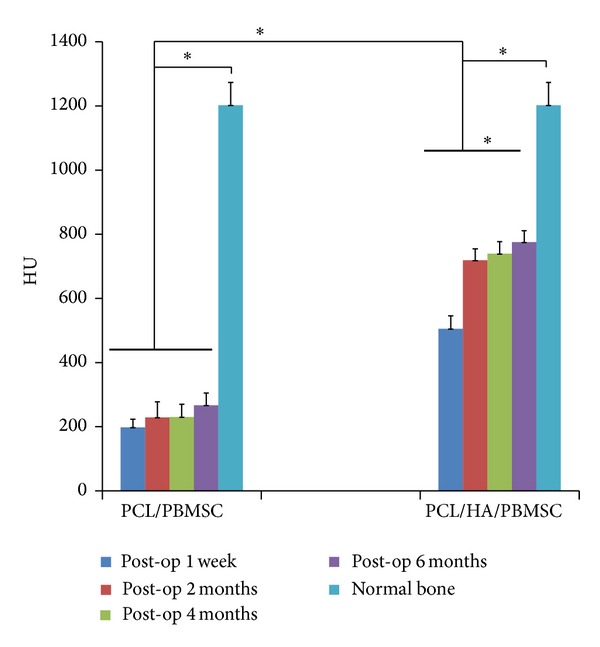
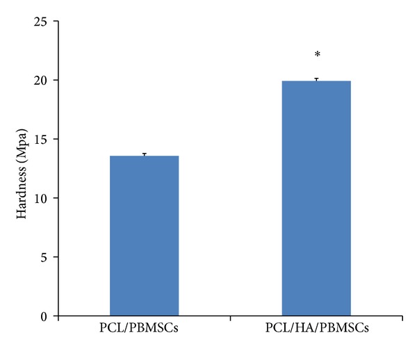
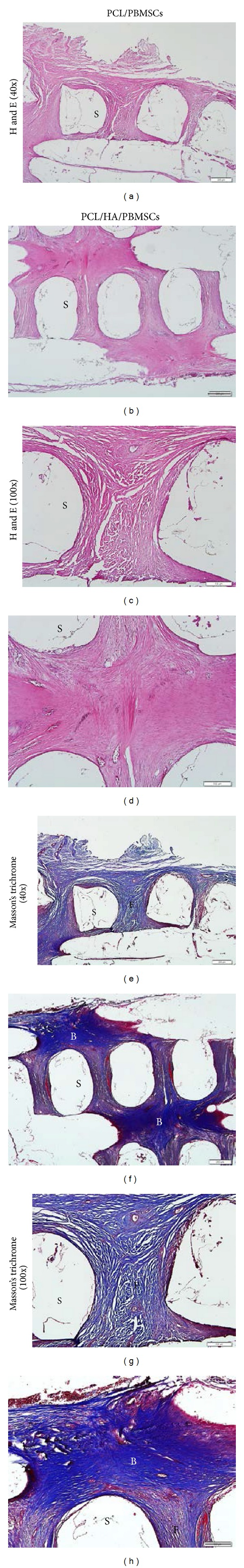
The study described a novel bone tissue scaffold fabricated by computer-aided, air pressure-aided deposition system to control the macro- and microstructure precisely. The porcine bone marrow stem cells (PBMSCs) seeded on either mPEG-PCL-mPEG (PCL) or mPEG-PCL-mPEG/hydroxyapatite (PCL/HA) composite scaffold were cultured under osteogenic medium to test the ability of osteogenesis in vitro. The experimental outcomes indicated that both scaffolds possessed adequate pore size, porosity, and hydrophilicity for the attachment and proliferation of PBMSCs and the PBMSCs expressed upregulated genes of osteogensis and angiogenesis in similar manner on both scaffolds. The major differences between these two types of the scaffolds were the addition of HA leading to higher hardness of PCL/HA scaffold, cell proliferation, and VEGF gene expression in PCL/HA scaffold. However, the in vivo bone forming efficacy between PBMSCs seeded PCL and PCL/HA scaffold was different from the in vitro results. The outcome indicated that the PCL/HA scaffold which had bone-mimetic environment due to the addition of HA resulted in better bone regeneration and mechanical strength than those of PCL scaffold. Therefore, providing a bone-mimetic scaffold is another crucial factor for bone tissue engineering in addition to the biocompatibility, 3D architecture with high porosity, and interpored connection.
Figures
Similar articles
-
Fabrication techniques involved in developing the composite scaffolds PCL/HA nanoparticles for bone tissue engineering applications.J Mater Sci Mater Med. 2021 Aug 11;32(8):93. doi: 10.1007/s10856-021-06564-0. J Mater Sci Mater Med. 2021. PMID: 34379204 Free PMC article. Review.
-
Selective laser sintering fabrication of nano-hydroxyapatite/poly-ε-caprolactone scaffolds for bone tissue engineering applications.Int J Nanomedicine. 2013;8:4197-213. doi: 10.2147/IJN.S50685. Epub 2013 Nov 1. Int J Nanomedicine. 2013. PMID: 24204147 Free PMC article.
-
Solid free-form fabrication-based PCL/HA scaffolds fabricated with a multi-head deposition system for bone tissue engineering.J Biomater Sci Polym Ed. 2010;21(6-7):951-62. doi: 10.1163/156856209X458380. J Biomater Sci Polym Ed. 2010. PMID: 20482995
-
Biofabrication and in vitro study of hydroxyapatite/mPEG-PCL-mPEG scaffolds for bone tissue engineering using air pressure-aided deposition technology.Mater Sci Eng C Mater Biol Appl. 2013 Mar 1;33(2):680-90. doi: 10.1016/j.msec.2012.10.018. Epub 2012 Nov 10. Mater Sci Eng C Mater Biol Appl. 2013. PMID: 25427474
-
Electropsun Polycaprolactone Fibres in Bone Tissue Engineering: A Review.Mol Biotechnol. 2021 May;63(5):363-388. doi: 10.1007/s12033-021-00311-0. Epub 2021 Mar 10. Mol Biotechnol. 2021. PMID: 33689142 Review.
Cited by
-
Fabrication techniques involved in developing the composite scaffolds PCL/HA nanoparticles for bone tissue engineering applications.J Mater Sci Mater Med. 2021 Aug 11;32(8):93. doi: 10.1007/s10856-021-06564-0. J Mater Sci Mater Med. 2021. PMID: 34379204 Free PMC article. Review.
-
Recent Advances in Bioengineering Bone Revascularization Based on Composite Materials Comprising Hydroxyapatite.Int J Mol Sci. 2023 Aug 6;24(15):12492. doi: 10.3390/ijms241512492. Int J Mol Sci. 2023. PMID: 37569875 Free PMC article. Review.
-
Fabrication and Application of a 3D-Printed Poly-ε-Caprolactone Cage Scaffold for Bone Tissue Engineering.Biomed Res Int. 2020 Jan 30;2020:2087475. doi: 10.1155/2020/2087475. eCollection 2020. Biomed Res Int. 2020. PMID: 32083125 Free PMC article.
-
Healing of Osteochondral Defects Implanted with Biomimetic Scaffolds of Poly(ε-Caprolactone)/Hydroxyapatite and Glycidyl-Methacrylate-Modified Hyaluronic Acid in a Minipig.Int J Mol Sci. 2018 Apr 9;19(4):1125. doi: 10.3390/ijms19041125. Int J Mol Sci. 2018. PMID: 29642550 Free PMC article.
-
Overexpression of VEGF in dermal fibroblast cells accelerates the angiogenesis and wound healing function: in vitro and in vivo studies.Sci Rep. 2022 Nov 2;12(1):18529. doi: 10.1038/s41598-022-23304-8. Sci Rep. 2022. PMID: 36323953 Free PMC article.
References
-
- Burg KJ, Porter S, Kellam JF. Biomaterial developments for bone tissue engineering. Biomaterials. 2000;21(23):2347–2359. - PubMed
-
- Yang S, Leong KF, Du Z, Chua CK. The design of scaffolds for use in tissue engineering. Part I. Traditional factors. Tissue Engineering. 2001;7(6):679–689. - PubMed
-
- Yang S, Leong KF, Du Z, Chua CK. The design of scaffolds for use in tissue engineering. Part II. Rapid prototyping techniques. Tissue Engineering. 2002;8(1):1–11. - PubMed
-
- Yeong WY, Chua CK, Leong KF, Chandrasekaran M. Rapid prototyping in tissue engineering: challenges and potential. Trends in Biotechnology. 2004;22(12):643–652. - PubMed
-
- Sachlos E, Czernuszka JT. Making tissue engineering scaffolds work. Review: the application of solid freeform fabrication technology to the production of tissue engineering scaffolds. European Cells & Materials. 2003;5:29–39. - PubMed
Publication types
MeSH terms
Substances
LinkOut - more resources
Full Text Sources
Other Literature Sources
Medical
