. 2014:2014:827327.
doi: 10.1155/2014/827327. Epub 2014 Apr 24.

Tumor and endothelial cell hybrids participate in glioblastoma vasculature

Affiliations

Tumor and endothelial cell hybrids participate in glioblastoma vasculature

Soufiane El Hallani et al. Biomed Res Int. 2014.

Abstract

Background: Recently antiangiogenic therapy with bevacizumab has shown a high but transient efficacy in glioblastoma (GBM). Indeed, GBM is one of the most angiogenic human tumors and endothelial proliferation is a hallmark of the disease. We therefore hypothesized that tumor cells may participate in endothelial proliferation of GBM.

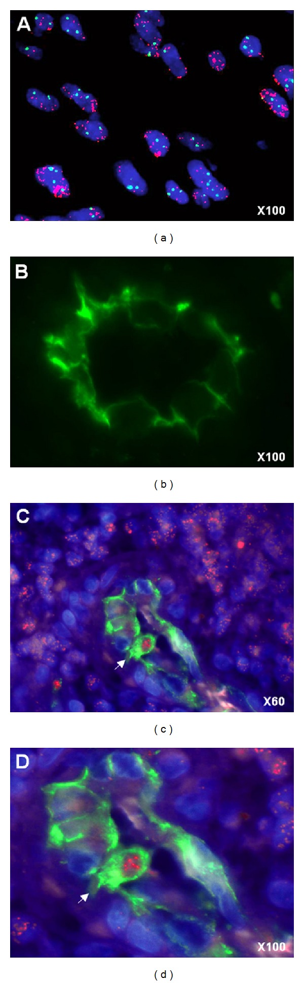
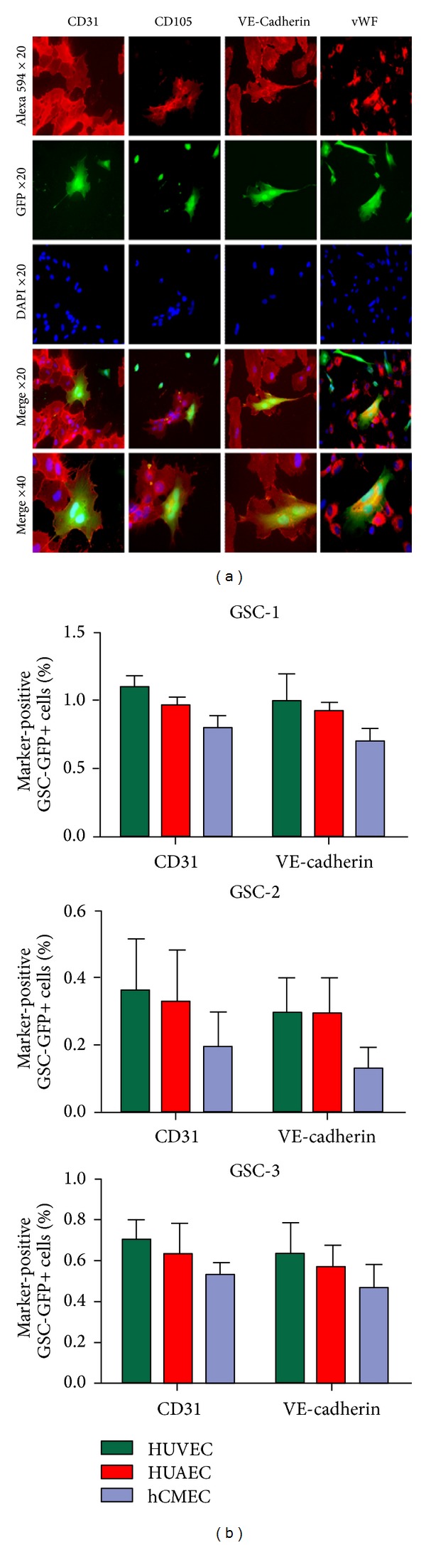
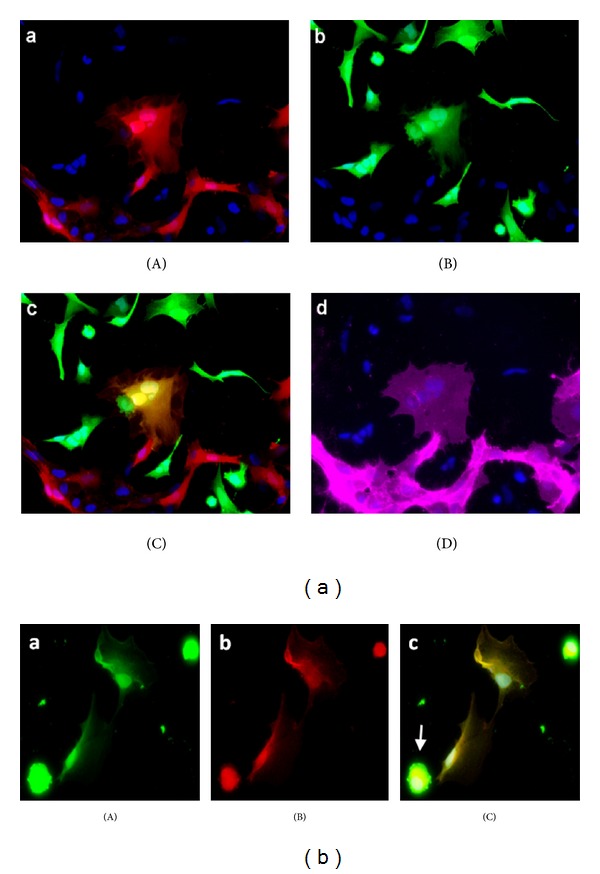
Materials and methods: We used EGFR FISH Probe to detect EGFR amplification and anti-CD31, CD105, VE-cadherin, and vWF to identify endothelial cells. Endothelial and GBM cells were grown separately, labeled with GFP and DsRed lentiviruses, and then cocultured with or without contact.

Results: In a subset of GBM tissues, we found that several tumor endothelial cells carry EGFR amplification, characteristic of GBM tumor cells. This observation was reproduced in vitro: when tumor stem cells derived from GBM were grown in the presence of human endothelial cells, a fraction of them acquired endothelial markers (CD31, CD105, VE-cadherin, and vWF). By transduction with GFP and DsRed expressing lentiviral vectors, we demonstrate that this phenomenon is due to cell fusion and not transdifferentiation.

Conclusion: A fraction of GBM stem cells thus has the capacity to fuse with endothelial cells and the resulting hybrids may participate in tumor microvascular proliferation and in treatment resistance.


Figures

Figure 1
Figure 1
Glioblastoma-derived endothelial cells. (a) Nuclei are stained with DAPI (blue), FISH EGFR probe (red) label tumor cells carrying EGFR amplification as double minutes (display multiple red signals). (b) Endothelial cells are detected by anti-CD31 immunofluorescence staining. ((c), (d)) A CD31+ (green) endothelial cell (arrow) carrying EGFR amplification (multiple red signals) is visible in a GBM microvessel.
Figure 2
Figure 2
Glioblastoma stem cell characterization. (a) Phase contrast and fluorescent microscopy of GSC-1-GFP+ growing into tumor spheres in neurosphere medium (magnification ×10). (b) Comparative genomic hybridization array (CGHa) of GSC-1 demonstrating tumor genomic alterations. Each BAC (bacterial artificial chromosome) spotted on the comparative genomic hybridization array is represented by a dot. BACs are ordered on the x-axis according to their position in the genome. For each chromosome, the telomere of the short arm is on the left and the telomere of the long arm is on the right. The y-axis corresponds to fluorescence ratio. Yellow, green, and red indicate, respectively, genomic copy number normal, loss, and gain. Genetic alteration includes complete chromosome 10 loss (green) and gain of chromosome 7 with EGFR amplification (arrow). (c) RT-PCR analysis showing specific expression of endothelial markers CD31 and VE-cadherin by hCMEC and not by GSC-1, GSC-2, and GSC-3 after differentiation in EGM-2 medium. ALAS is used as control. (d) Immunostaining of tumor sphere cells for neural stem cell marker (nestin) at the beginning of the differentiation assay, then astrocytic (GFAP) and neuronal (TUJ-1) markers by the differentiated cells around a tumor sphere at day 7 (magnification ×10).
Figure 3
Figure 3
A subset of glioblastoma stem cells acquires an endothelial phenotype. (a) When GSC-GFP were cocultured with human endothelial cells, several multinucleated and cobblestone GSC-GFP expressing CD31, CD105, VE-cadherin, and vWF were observed (b). The percentage of GSC-GFP expressing CD31 and VE-cadherin obtained in coculture with HUVEC, HUAEC, and hCMEC is indicated for GSC-1, GSC-2, and GSC-3.
Figure 4
Figure 4
Glioblastoma stem cell fusion with endothelial cells. (a) When GSC-GFP were cocultured with HUVEC-DsRed, we observed binucleated cell expressing both DsRed (A) and GFP (B) (merged in (C)) corresponding to hybrid cell and exhibiting endothelial phenotype as shown by CD31 immunostaining (D). (b) After hybrid cell selection by cell sorting, mononuclear hybrids expressing parental GFP (A) and DsRed (B) were observed (merged in (C)). Apoptotic body of died fused cell (arrow).

References

    1. Louis DN, Ohgaki H, Wiestler OD, et al. The 2007 WHO classification of tumours of the central nervous system. Acta Neuropathologica. 2007;114(2):97–109. - PMC - PubMed
    1. Vredenburgh JJ, Desjardins A, Herndon JE, II, et al. Phase II trial of bevacizumab and irinotecan in recurrent malignant glioma. Clinical Cancer Research. 2007;13(4):1253–1259. - PubMed
    1. Kreisl TN, Kim L, Moore K, et al. Phase II trial of single-agent bevacizumab followed by bevacizumab plus irinotecan at tumor progression in recurrent glioblastoma. Journal of Clinical Oncology. 2009;27(5):740–745. - PMC - PubMed
    1. McDonald DM, Choyke PL. Imaging of angiogenesis: from microscope to clinic. Nature Medicine. 2003;9(6):713–725. - PubMed
    1. Mollica F, Jain RK, Netti PA. A model for temporal heterogeneities of tumor blood flow. Microvascular Research. 2003;65(1):56–60. - PubMed

Publication types

MeSH terms