

Current concepts and future directions for the assessment of autoantibodies to cellular antigens referred to as anti-nuclear antibodies
- PMID: 24868563
- PMCID: PMC4020446
- DOI: 10.1155/2014/315179
Current concepts and future directions for the assessment of autoantibodies to cellular antigens referred to as anti-nuclear antibodies
Abstract
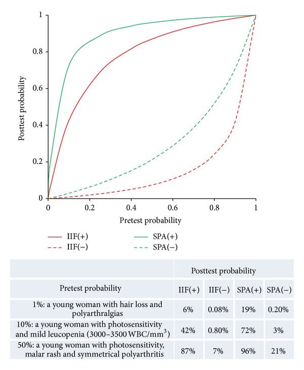
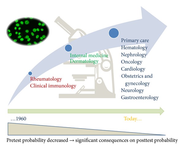
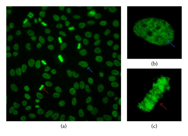
The detection of autoantibodies that target intracellular antigens, commonly termed anti-nuclear antibodies (ANA), is a serological hallmark in the diagnosis of systemic autoimmune rheumatic diseases (SARD). Different methods are available for detection of ANA and all bearing their own advantages and limitations. Most laboratories use the indirect immunofluorescence (IIF) assay based on HEp-2 cell substrates. Due to the subjectivity of this diagnostic platform, automated digital reading systems have been developed during the last decade. In addition, solid phase immunoassays using well characterized antigens have gained widespread adoption in high throughput laboratories due to their ease of use and open automation. Despite all the advances in the field of ANA detection and its contribution to the diagnosis of SARD, significant challenges persist. This review provides a comprehensive overview of the current status on ANA testing including automated IIF reading systems and solid phase assays and suggests an approach to interpretation of results and discusses meeting the problems of assay standardization and other persistent challenges.
Figures
References
-
- Friou GJ, Finch SC, Detre KD. Interaction of nuclei and globulin from lupus erythematosis serum demonstrated with fluorescent antibody. Journal of Immunology. 1958;80(4):324–329. - PubMed
-
- Friou GJ. Setting the scene: a historical and personal view of immunologic diseases, autoimmunity and ANA. Clinical and Experimental Rheumatology. 1994;12(supplement 11):S23–S25. - PubMed
-
- Hargraves MM, Richmond H, Morton R. Presentation of two bone marrow elements, the tart cell and the L.E. cell. Proceedings of the Staff Meetings of the Mayo Clinic. 1948;23:25–28. - PubMed
Publication types
MeSH terms
Substances
LinkOut - more resources
Full Text Sources
Other Literature Sources
Medical
