. 2014 May;243(5):730-8.
doi: 10.1002/dvdy.24105.

Expression of actin-binding proteins and requirement for actin-depolymerizing factor in chick neural crest cells

Expression of actin-binding proteins and requirement for actin-depolymerizing factor in chick neural crest cells

Katie L Vermillion et al. Dev Dyn. 2014 May.

Abstract

Background: Neural crest cells are multipotent cells that migrate extensively throughout vertebrate embryos to form diverse lineages. Cell migration requires polarized, organized actin networks that provide the driving force for motility. Actin-binding proteins that regulate neural crest cell migration are just beginning to be defined.

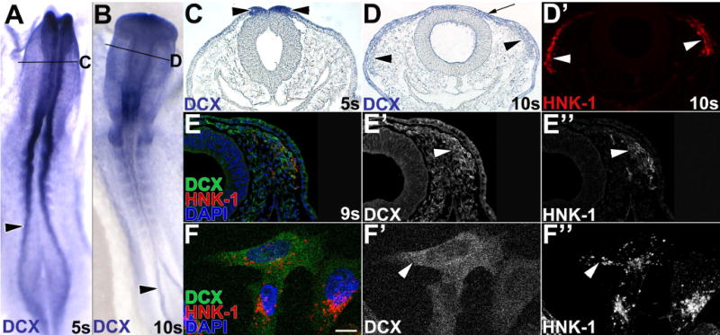
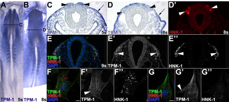
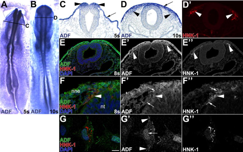
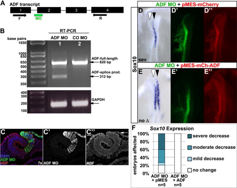
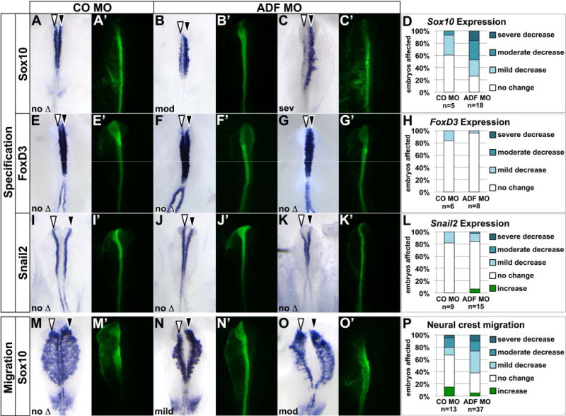
Results: We recently identified a number of actin-associated factors through proteomic profiling of methylated proteins in migratory neural crest cells. Here, we report the previously undocumented expression pattern of three of these proteins in chick early neural crest development: doublecortin (DCX), tropomyosin-1 (TPM-1), and actin depolymerizing factor (ADF). All three genes are expressed with varying degrees of specificity and intensity in premigratory and migratory neural crest cells, and their resulting proteins exhibit distinct subcellular localization in migratory neural crest cells. Morpholino knock down of ADF reveals it is required for Sox10 gene expression, but minimally important during neural crest migration.

Conclusions: Neural crest cells express DCX, TPM-1, and ADF. ADF is necessary during neural crest specification, but largely dispensable for migration.


Figures

Figure 1
Figure 1. Doublecortin is expressed throughout neural crest development
(A–D) Premigratory and migratory neural crest cells express DCX mRNA. DCX expression was visualized in 5 somite (s; A,C) and 10s (B,D) chick embryos by in situ hybridization. Sections (C,D) were taken at the level indicated in the whole mount view (A,B). At 5s, DCX mRNA is broadly expressed, but particularly abundant in neural folds (black arrowheads) in whole mount (A) and cross-section (C). At 10s in whole mount (B), DCX is highest cranially and in developing somites and caudal neural folds. In a midbrain cross-section at 10s (D), DCX mRNA is expressed in non-neural ectoderm (arrow), and in HNK-1-positive (D′, white arrowheads) migratory neural crest cells (black arrowheads). A,B, dorsal view. (E–F) DCX protein is expressed in migratory neural crest cells by immunofluorescence. In a 9 somite (E–E″) chick embryo midbrain section, DCX protein is abundant in cranial migratory neural crest cells (E, green; E′, white arrowhead) positive for HNK-1 (E, red; E″, white arrowhead), and expressed at lower levels in non-neural ectoderm and head mesenchyme. In cultured cranial migratory neural crest cells (F), DCX protein (F, green; F′, white arrowhead) is cytoplasmic and highest around the nucleus.
Figure 2
Figure 2. Tropomyosin 1 expression in the neural crest
(A–D) Neural crest cells express low-levels of TPM-1 mRNA. TPM-1 expression was visualized in 5 somite (s; A,C) and 8s (B,D) chick embryos by in situ hybridization. Sections (C,D) were taken at the level indicated in the whole mount view (A,B). In whole mount, TPM-1 mRNA is expressed everywhere except the midline and neural plate (A,B). In a midbrain sections, non-neural ectoderm and head mesenchyme strongly express TPM-1, while the neural tube does not (C,D). TPM-1 expression is moderate in neural folds (C, black arrowheads), but declines in HNK-1-positive (D′, white arrowhead) migratory neural crest cells moving away from the neural tube (D, black arrowheads). A,B, dorsal view. (E–G) Leading migratory neural crest cells contain the motility-activated TPM-1 epitope. In a midbrain section of a 9 somite (E–E″) chick embryo, immunofluorescence with the CG-1 antibody (Hegmann et al., 1988) shows motility-dependent TPM-1 immunoreactivity in cranial migratory neural crest cells (E, green, E′, white arrowhead) that are positive for HNK-1 (E″, white arrowhead). HNK-1 positive (F,G, red; F″, G″) cultured cranial migratory neural crest cells exhibit motility-activated TPM-1 immunoreactivity (F,G, green; F′, G′) in striped patterns throughout the cell body.
Figure 3
Figure 3. Premigratory and migratory neural crest cells express ADF
(A–D) Neural crest cells express ADF mRNA. In situ hybridization for ADF in 5 somite (s; A,C) and 10s (B,D) chick embryos. Sections (C–D) were taken at the level indicated in the whole mount view (A,B). In whole mount, ADF mRNA is widely expressed (A–B). In sections, ADF is particularly abundant in premigratory neural crest cells in the neural folds (C, black arrowheads) and in HNK-1-positive (D′, white arrowhead) migratory neural crest cells (D, black arrowheads). Non-neural ectoderm (C,D, arrow) and the basal surface of the neural tube also strongly express ADF. (E–G) Migratory neural crest cells express ADF protein. In 8 somite chick embryo midbrain sections (E–F), immunofluorescence reveals abundant ADF protein (E,F, green; E′,F′) in the non-neural ectoderm (nne) and HNK-1-positive (E,F red; E″,F″ white arrowheads) cranial migratory neural crest cells. ADF is both cytoplasmic (Fig. 3F′, arrowhead) and nuclear (Fig. 3F′ white arrows). In cultured cranial neural neural crest cells (G–G″). ADF protein (G, green; G′, white arrowheads) is nuclear (G′, white arrow), along the cell periphery, and at the tips of protrusions of HNK-1-positive (G, red; G″) migratory neural crest cells (G′, arrowheads).
Figure 4
Figure 4. ADF MO elicits ADF-specific knock down
(A) ADF exon (numbered rectangles) and intron (lines) organization, splice-blocking MO (green bar) site, and forward (F) and reverse (R) primer (arrows) locations. (B,C) Embryos were unilaterally electroporated with ADF MO (B, lane 1; C, green; C′) or standard control MO (CO MO; B, lane 2) at late gastrula, reincubated to 7 somites (7s), and RT-PCR (B) or ADF immunofluorescence (C) performed. ADF MO promotes ADF mis-splicing (B, lane 1; 312 bp (base pair) product), while CO MO yields normally spliced ADF (B, lane 2; 620 bp product). GAPDH shows equal input cDNA. Meanwhile, in ADF MO-electroporated embryo sections, ADF immunoreactivity (C, red; C″) is reduced in ADF MO-targeted cells (C, green; C′; C″, white arrow) compared to untargeted cells (C, green; C″, black arrow). (D–F) ADF co-expression rescues ADF knock down. Embryos were unilaterally electroporated with ADF MO (D′,E′, green) mixed with pMES-mCherry (D″, red) or pMES-mCherry-ADF (E″, red) at late gastrula, reincubated to 4–6 somites, and Sox10 visualized by in situ hybridization (D,E, purple; dorsal view). White arrowhead, targeted side; black arrowhead, untargeted side. (F) Stacked bar graph shows that ADF co-expression rescues ADF MO-dependent loss of Sox10 expression.
Fig. 5
Fig. 5. ADF is required for neural crest specification and migration
Embryos were unilaterally electroporated with standard control MO (CO MO; A,E,I,M, green) or ADF MO (B,C,F,G,J,K,N,O, green) at late gastrula, reincubated to 4–6 somites (A–C, E–G, I–K) or 8–10 somites (M–O), and processed by in situ hybridization (purple) to visualize expression of Sox10 (A–C, M–O), FoxD3 (E–G), or Snail2 (I–K). Dorsal view of in situ hybridization in left panel, fluorescent MO targeting in right panel. White arrowhead, targeted side of embryo; black arrowhead, untargeted side of embryo. (A–J) ADF is necessary for Sox10, but not FoxD3 or Snail2 expression. (A–C, E–G, I–K) Representative examples of electroporated embryos, showing only Sox10 expression defects on the ADF MO-targeted side. (D,H,L) Stacked bar graph depicting the frequency and severity of gene expression defects in embryos electroporated with CO MO or ADF MO. (M–P) ADF knockdown causes subtle defects in neural crest migration distance. (M–O) Representative examples of electroporated embryos, with the majority of ADF-targeted embryos mildly affected. (N) Stacked bar graph depicting the frequency and severity of migration defects in embryos electroporated with CO MO or ADF MO.

Similar articles

Cited by

References

    1. Abe H, Ohshima S, Obinata T. A cofilin-like protein is involved in the regulation of actin assembly in developing skeletal muscle. J Biochem. 1989;106:696–702. - PubMed
    1. Bach CT, Creed S, Zhong J, Mahmassani M, Schevzov G, Stehn J, Cowell LN, Naumanen P, Lappalainen P, Gunning PW, O’Neill GM. Tropomyosin isoform expression regulates the transition of adhesions to determine cell speed and direction. Mol Cell Biol. 2009;29:1506–1514. - PMC - PubMed
    1. Baker JC, Beddington RS, Harland RM. Wnt signaling in Xenopus embryos inhibits bmp4 expression and activates neural development. Genes Dev. 1999;13:3149–3159. - PMC - PubMed
    1. Bamburg JR, Bray D. Distribution and cellular localization of actin depolymerizing factor. J Cell Biol. 1987;105:2817–2825. - PMC - PubMed
    1. Bamburg JR, Wiggan OP. ADF/cofilin and actin dynamics in disease. Trends Cell Biol. 2002;12:598–605. - PubMed

Publication types

MeSH terms