. 2014 May 28;9(5):e98480.
doi: 10.1371/journal.pone.0098480. eCollection 2014.

Accelerated postero-lateral spinal fusion by collagen scaffolds modified with engineered collagen-binding human bone morphogenetic protein-2 in rats

Affiliations

Accelerated postero-lateral spinal fusion by collagen scaffolds modified with engineered collagen-binding human bone morphogenetic protein-2 in rats

Xinglong Han et al. PLoS One. .

Abstract

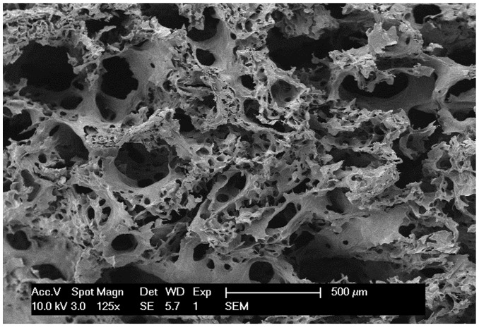
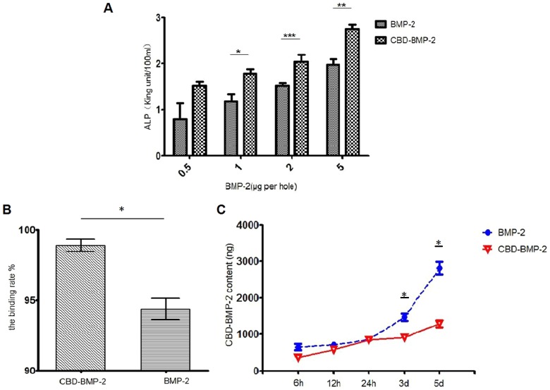
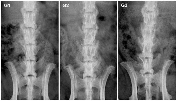
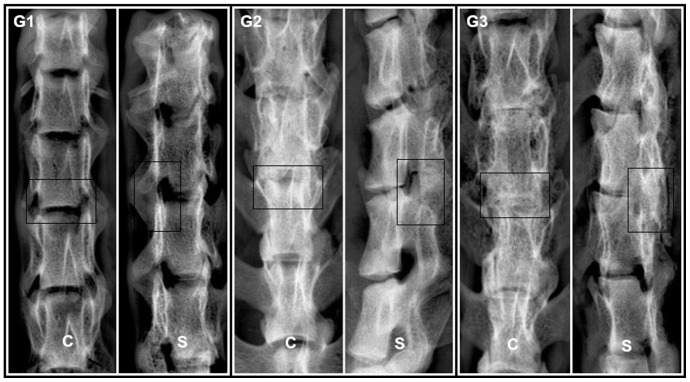
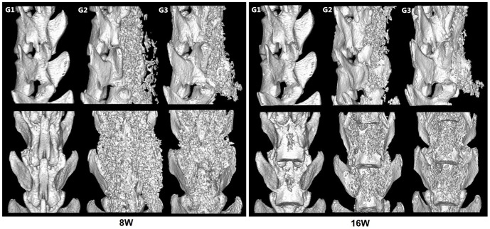
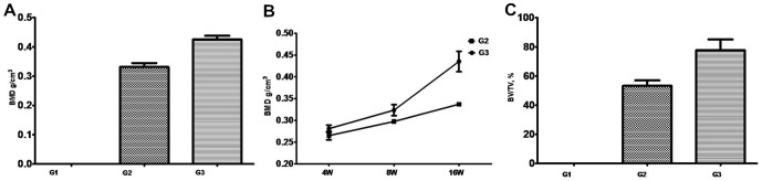
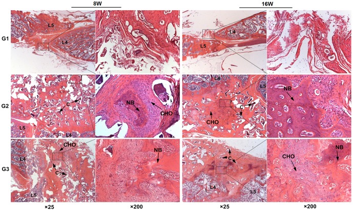
Bone morphogenetic protein-2 (BMP-2) is a potent osteoinductive cytokine that plays a critical role in bone regeneration and repair. However, its distribution and side effects are major barriers to its success as therapeutic treatment. The improvement of therapy using collagen delivery matrices has been reported. To investigate a delivery system on postero-lateral spinal fusion, both engineered human BMP-2 with a collagen binding domain (CBD-BMP-2) and collagen scaffolds were developed and their combination was implanted into Sprague-Dawley (SD) rats to study Lumbar 4-5 (L4-L5) posterolateral spine fusion. We divided SD rats into three groups, the sham group (G1, n = 20), the collagen scaffold-treated group (G2, n = 20) and the BMP-2-loaded collagen scaffolds group (G3, n = 20). 16 weeks after surgery, the spines of the rats were evaluated by X-radiographs, high-resolution micro-computed tomography (micro-CT), manual palpation and hematoxylin and eosin (H&E) staining. The results showed that spine L4-L5 fusions occurred in G2(40%) and G3(100%) group, while results from the sham group were inconsistent. Moreover, G3 had better results than G2, including higher fusion efficiency (X score, G2 = 2.4±0.163, G3 = 3.0±0, p<0.05), higher bone mineral density (BMD, G2: 0.3337±0.0025g/cm3, G3: 0.4353±0.0234g/cm3. p<0.05) and more bone trabecular formation. The results demonstrated that with site-specific collagen binding domain, a dose of BMP-2 as low as 0.02mg CBD-BMP-2/cm3 collagen scaffold could enhance the posterolateral intertransverse process fusion in rats. It suggested that combination delivery could be an alternative in spine fusion with dramatically decreased side effects caused by high dose of BMP-2.


Conflict of interest statement

Competing Interests: The authors have declared that no competing interests exist.

Figures

Figure 1
Figure 1. The morphology of the processed collagen scaffolds.
This granular scaffolds were taken photographs under the scanning electron microscope (125×). The micropores size range from 100 µm to 300 µm.
Figure 2
Figure 2. The release profile and bioactivity assay of BMP-2 in vitro.
A: The CBD-BMP-2 shows a higher biological activity than the commercial BMP-2 (*:p<0.05; **:p<0.01; ***:p<0.001;). B: The binding rate of CBD-BMP-2 to the collagen scaffolds is higher than that of commercial BMP-2(p<0.05). C: The releasing amount and rate in the commercial BMP-2 group was higher than those of the CBD-BMP-2 group.(*, Compare to the control group, p<0.05).
Figure 3
Figure 3. X-Radiographs of SD rat spines in vivo.
Representative images of rats in each group are shown (coronal view). 8 weeks after the implantation, the spines appeared fused in G3, partially fused in G2 and no fused in G1.
Figure 4
Figure 4. X-radiographs of SD rat spines in vitro.
The Representative images showed that visible fusion occurred in both G2 and G3 groups at 16 weeks after the implantation (coronal view (C) and sagittal view (S)). The intervertebral space in G3 was hid under the new bone. A great quantity of new bone could be found and the processus transversus were joined to form a whole from a posterior lateral aspect between L4 and L5. There was a similar, but slightly imperfect result in G2.
Figure 5
Figure 5. Representative 3D reconstruction images of Micro-CT images.
8W: The images of 8 weeks after the implantation showed that obvious new bone formed in G2 and G3 while a similar phenomenon did not appear in G1. 16W: The images of 16weeks after the implantation showed that the spines of all the rats exhibited considerable new bone formation in G3 and fusion masses were significantly larger and denser than G2. There was still no new bone found in G1.
Figure 6
Figure 6. Bone mineral density (BMD) analysis by Micro-CT.
A: BMD analysis at 16 weeks after surgery. B: The BMD at different time points. C: The ratio of new bone volume (BV) to tissue volume (TV) at 16weeks. Data are expressed as the mean±SD (n = 10 per each group).
Figure 7
Figure 7. The H&E staining for L4–L5 posterolateral space in rats.
There were plenty of trabeculae between spongy bone which mean a fusion in G2 and G3 and no new bone were found in G1 (C: incompletely degraded collagen scaffolds, CHO: chondrocyte, NB: new bone).

References

    1. Reid JJ, Johnson JS, Wang JC (2011) Challenges to bone formation in spinal fusion. J Biomech 44: 213–220. - PubMed
    1. Kettler A, Rohlmann F, Ring C, Mack C, Wilke HJ (2011) Do early stages of lumbar intervertebral disc degeneration really cause instability? Evaluation of an in vitro database. Eur Spine J 20: 578–584. - PMC - PubMed
    1. Alsaleh KA, Tougas CA, Roffey DM, Wai EK (2012) Osteoconductive bone graft extenders in posterolateral thoracolumbar spinal fusion. Spine(Phila Pa 1976) 37: 993–1000. - PubMed
    1. Geiger M, Li RH, Friess W (2003) Collagen sponges for bone regeneration with rhBMP-2. Adv Drug Deliv Rev 55: 1613–1629. - PubMed
    1. Dewan AK, Dewan RA, Calderon N, Fuentes A, Lazard Z, et al. (2010) Assessing mechanical integrity of spinal fusion by in situ endochondral osteoinduction in the murine model. J Orthop Surg Res 5: 58–67. - PMC - PubMed

Publication types