

Near-infrared low-level laser stimulation of telocytes from human myometrium
- PMID: 24870411
- PMCID: PMC4215113
- DOI: 10.1007/s10103-014-1589-1
Near-infrared low-level laser stimulation of telocytes from human myometrium
Abstract
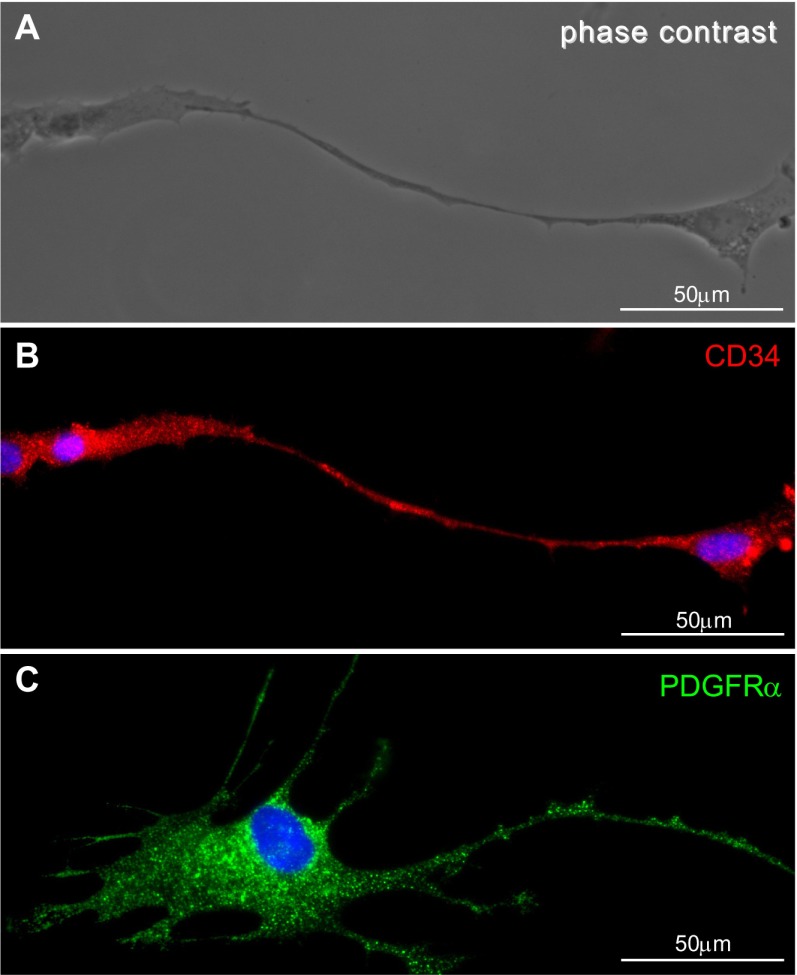
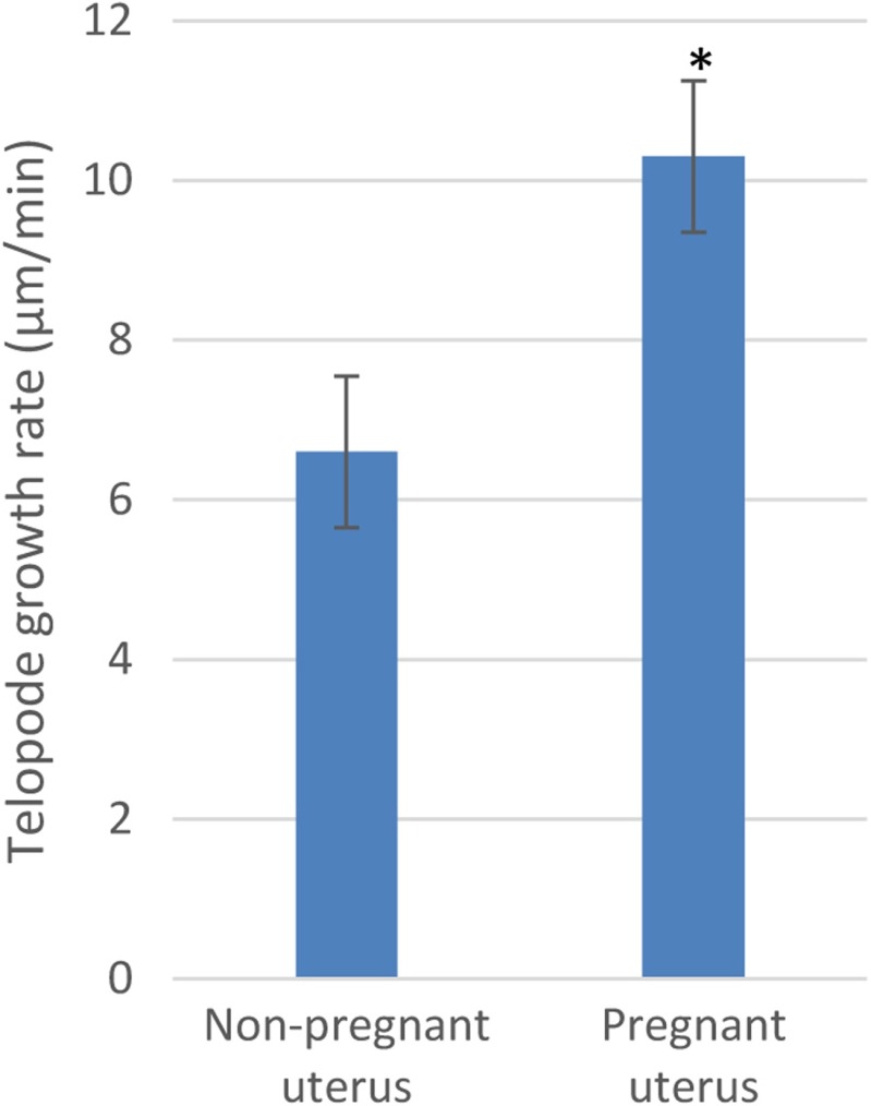
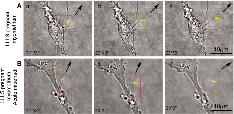
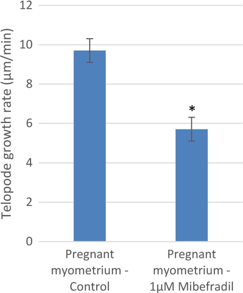
Telocytes (TCs) are a brand-new cell type frequently observed in the interstitial space of many organs (see www.telocytes.com ). TCs are defined by very long (tens of micrometers) and slender prolongations named telopodes. At their level, dilations-called podoms (~300 nm), alternate with podomers (80-100 nm). TCs were identified in a myometrial interstitial cell culture based on morphological criteria and by CD34 and PDGF receptor alpha (PDGFRα) immunopositivity. However, the mechanism(s) of telopodes formation and/or elongation and ramification is not known. We report here the low-level laser stimulation (LLLS) using a 1,064-nm neodymium-doped yttrium aluminum garnet (Nd:YAG) laser (with an output power of 60 mW) of the telopodal lateral extension (TLE) growth in cell culture. LLLS of TCs determines a higher growth rate of TLE in pregnant myometrium primary cultures (10.3 ± 1.0 μm/min) compared to nonpregnant ones (6.6 ± 0.9 μm/min). Acute exposure (30 min) of TCs from pregnant myometrium to 1 μM mibefradil, a selective inhibitor of T-type calcium channels, determines a significant reduction in the LLLS TLE growth rate (5.7 ± 0.8 μm/min) compared to LLLS per se in same type of samples. Meanwhile, chronic exposure (24 h) completely abolishes the LLLS TLE growth in both nonpregnant and pregnant myometria. The initial direction of TLE growth was modified by LLLS, the angle of deviation being more accentuated in TCs from human pregnant myometrium than in TCs from nonpregnant myometrium. In conclusion, TCs from pregnant myometrium are more susceptible of reacting to LLLS than those from nonpregnant myometrium. Therefore, some implications are emerging for low-level laser therapy (LLLT) in uterine regenerative medicine.
Figures

Similar articles
-
Telopodes of telocytes are influenced in vitro by redox conditions and ageing.Mol Cell Biochem. 2015 Dec;410(1-2):165-74. doi: 10.1007/s11010-015-2548-2. Epub 2015 Sep 3. Mol Cell Biochem. 2015. PMID: 26335900
-
Beta-Estradiol Regulates Voltage-Gated Calcium Channels and Estrogen Receptors in Telocytes from Human Myometrium.Int J Mol Sci. 2018 May 9;19(5):1413. doi: 10.3390/ijms19051413. Int J Mol Sci. 2018. PMID: 29747396 Free PMC article.
-
Isolated human uterine telocytes: immunocytochemistry and electrophysiology of T-type calcium channels.Histochem Cell Biol. 2015 Jan;143(1):83-94. doi: 10.1007/s00418-014-1268-0. Epub 2014 Sep 12. Histochem Cell Biol. 2015. PMID: 25212658 Free PMC article.
-
The Cutaneous Telocytes.Adv Exp Med Biol. 2016;913:303-323. doi: 10.1007/978-981-10-1061-3_20. Adv Exp Med Biol. 2016. PMID: 27796896 Review.
-
Telocytes in skeletal, cardiac and smooth muscle interstitium: morphological and functional aspects.Histol Histopathol. 2018 Nov;33(11):1151-1165. doi: 10.14670/HH-11-994. Epub 2018 Apr 25. Histol Histopathol. 2018. PMID: 29693711 Review.
Cited by
-
FIB-SEM tomography of human skin telocytes and their extracellular vesicles.J Cell Mol Med. 2015 Apr;19(4):714-22. doi: 10.1111/jcmm.12578. J Cell Mol Med. 2015. PMID: 25823591 Free PMC article.
-
Estradiol enhances T-type calcium channel activation in human myometrium telocytes.J Reprod Dev. 2023 Apr 3;69(2):87-94. doi: 10.1262/jrd.2022-132. Epub 2023 Feb 9. J Reprod Dev. 2023. PMID: 36754390 Free PMC article.
-
Ultrastructural and immunohistochemical characteristics of telocytes in human scalp tissue.Sci Rep. 2020 Feb 3;10(1):1693. doi: 10.1038/s41598-020-58628-w. Sci Rep. 2020. PMID: 32015359 Free PMC article.
-
Telocytes in female reproductive system (human and animal).J Cell Mol Med. 2016 Jun;20(6):994-1000. doi: 10.1111/jcmm.12843. Epub 2016 Apr 5. J Cell Mol Med. 2016. PMID: 27060783 Free PMC article. Review.
-
Telocytes and Their Structural Relationships With the Sperm Storage Tube and Surrounding Cell Types in the Utero-Vaginal Junction of the Chicken.Front Vet Sci. 2022 Mar 24;9:852407. doi: 10.3389/fvets.2022.852407. eCollection 2022. Front Vet Sci. 2022. PMID: 35400114 Free PMC article.
References
-
- Mou Y, Wang Y, Li J, Lü S, Duan C, Du Z, Yang G, Chen W, Zhao S, Zhou J, Wang C. Immunohistochemical characterization and functional identification of mammary gland telocytes in the self-assembly of reconstituted breast cancer tissue in vitro. J Cell Mol Med. 2013;17:65–75. doi: 10.1111/j.1582-4934.2012.01646.x. - DOI - PMC - PubMed
Publication types
MeSH terms
Substances
LinkOut - more resources
Full Text Sources
Other Literature Sources
