. 2014 May 28;6(238):238ra71.
doi: 10.1126/scitranslmed.3008004.

Hematopoietic stem cell origin of BRAFV600E mutations in hairy cell leukemia

Affiliations

Hematopoietic stem cell origin of BRAFV600E mutations in hairy cell leukemia

Stephen S Chung et al. Sci Transl Med. .

Abstract

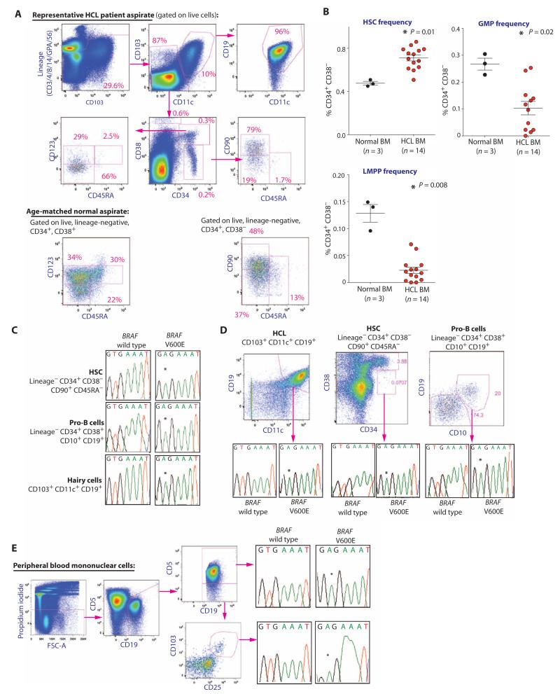
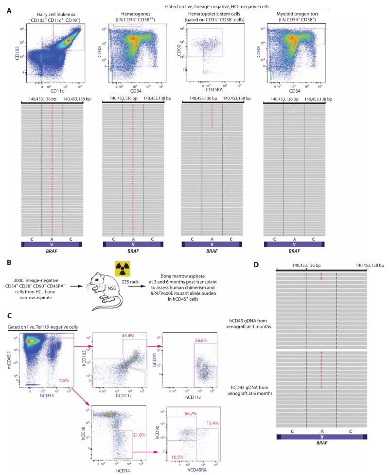
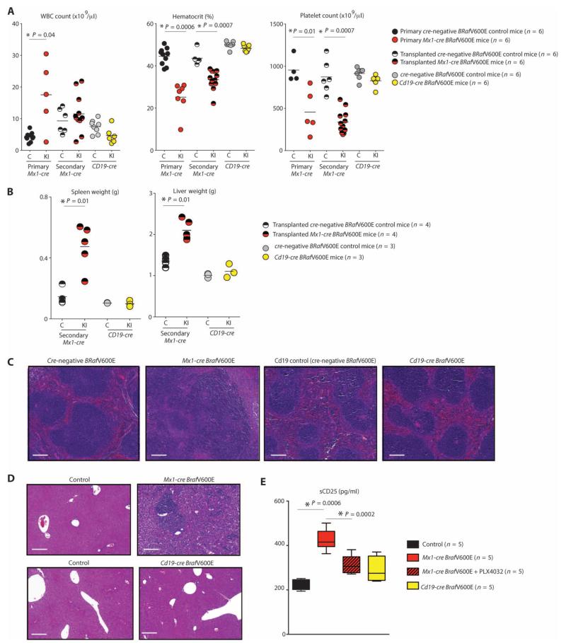
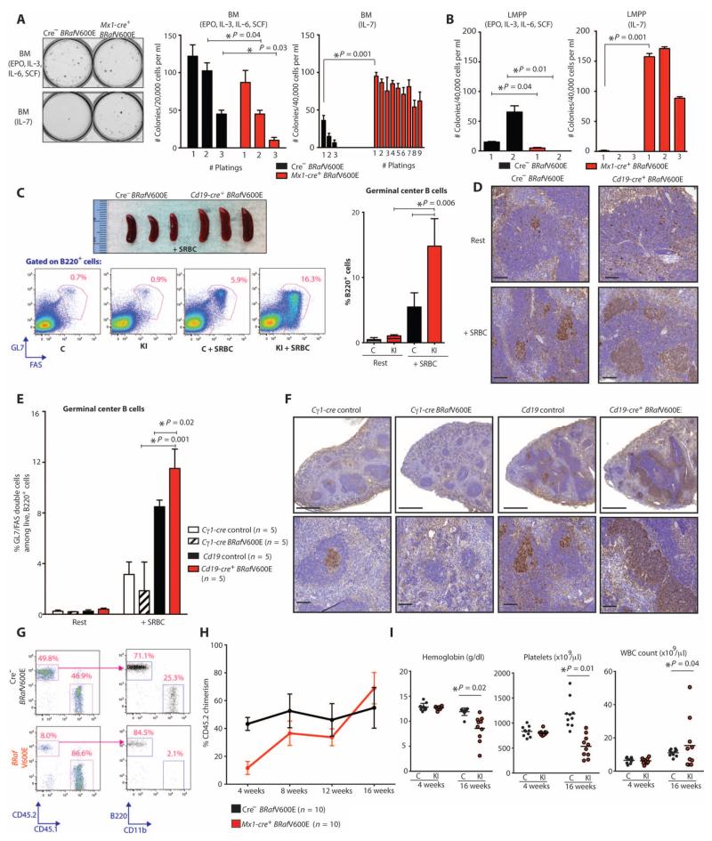
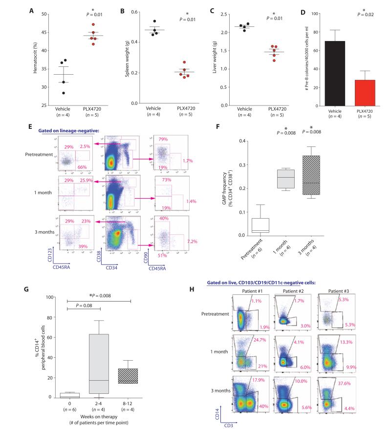
Hairy cell leukemia (HCL) is a chronic lymphoproliferative disorder characterized by somatic BRAFV600E mutations. The malignant cell in HCL has immunophenotypic features of a mature B cell, but no normal counterpart along the continuum of developing B lymphocytes has been delineated as the cell of origin. We find that the BRAFV600E mutation is present in hematopoietic stem cells (HSCs) in HCL patients, and that these patients exhibit marked alterations in hematopoietic stem/progenitor cell (HSPC) frequencies. Quantitative sequencing analysis revealed a mean BRAFV600E-mutant allele frequency of 4.97% in HSCs from HCL patients. Moreover, transplantation of BRAFV600E-mutant HSCs from an HCL patient into immunodeficient mice resulted in stable engraftment of BRAFV600E-mutant human hematopoietic cells, revealing the functional self-renewal capacity of HCL HSCs. Consistent with the human genetic data, expression of BRafV600E in murine HSPCs resulted in a lethal hematopoietic disorder characterized by splenomegaly, anemia, thrombocytopenia, increased circulating soluble CD25, and increased clonogenic capacity of B lineage cells-all classic features of human HCL. In contrast, restricting expression of BRafV600E to the mature B cell compartment did not result in disease. Treatment of HCL patients with vemurafenib, an inhibitor of mutated BRAF, resulted in normalization of HSPC frequencies and increased myeloid and erythroid output from HSPCs. These findings link the pathogenesis of HCL to somatic mutations that arise in HSPCs and further suggest that chronic lymphoid malignancies may be initiated by aberrant HSCs.


Figures

Fig. 1
Fig. 1. HSPC abnormalities and the presence of the BRAFV600E mutation in HSCs of HCL patients
(A) Stem and progenitor flow cytometric analysis of the BM of a representative HCL patient and an age-matched control. The sort schema shown was used for isolation of HCL cells (CD103+ CD19+ CD11c+ cells), HSCs (LN CD34+ CD38 CD90+ CD45RA cells), hematogones (CD34 CD38++ CD10+ CD19+), and MP cells (LN CD34+ CD38+ CD45RA+/ CD123+/ cells). (B) Frequencies of HSCs, LMPPs (LN CD34+ CD38 CD90 CD45RA+), and GMPs (LN CD34+ CD38+ CD123+ CD45RA+) in 14 patients with HCL and 3 age-matched normal control BM aspirate samples. (C) Prospective cell separation including double sorting to ensure purity and lack of HCL cell contamination followed by allele-specific polymerase chain reaction (PCR) analysis for the presence of the BRAFV600E mutation reveals the mutation in HSCs, pro-B cells, and HCL cells. (D) Similar data from a second HCL patient revealing the mutation in HSC, pro-B, and HCL double-sorted cell populations. (E) Prospective isolation of CLL (CD19+ CD5+ CD103 CD11c cells) and HCL cell populations (CD19+ CD103+ CD11c+ CD5 cells) from the PB of one individual with both disorders reveals the BRAFV600E mutation in both cell populations. Error bars represent means ± SD. *P < 0.05 (Mann-Whitney U test).
Fig. 2
Fig. 2. Quantitative analysis of the BRAFV600E mutation in HSCs from HCL patients and functional self-renewal capacity of BRAFV600E-mutant HSCs
(A) Representative FACS analysis and quantitative sequencing analysis revealing the VAF of the BRAF c.T1860A p.V600E mutation in HCL cells, hematogones, HSCs, and MP cells from an HCL patient (for clarity, only 52 reads are displayed). cDNA from double FACS-sorted cell populations were used for MiSeq targeted sequencing. (B) Schema of xenograft experiment where 3000 HSCs from a BRAFV600E-mutant HCL patient were injected into sublethally irradiated NSG mice followed by flow cytometric analysis of human engraftment and HCL cells, as well as quantification of the BRAFV600E mutation by sequencing analysis. (C) At 6 months after transplant, overall human chimerism was 4.5% with the presence of human HSCs (hCD45+ hCD34+ hCD38 hCD90+ hCD45RA) and a cell population with the immunophenotype of HCL cells (hCD45+ hCD103+ hCD19+ hCD11c+). (D) MiSeq sequencing analysis at 100× coverage reveals BRAFV600E mutation in 4 and 9% of hCD45+ cell genomic DNA at 3 and 6 months, respectively.
Fig. 3
Fig. 3. Phenotypic analysis of mice with pan-hematopoietic versus B lineage–restricted expression of BRafV600E
(A) White blood cell (WBC) count, hematocrit, and platelet count in 3-week-old primary Mx1-cre BRafV600E mice, lethally irradiated CD45.2 recipient mice 6 weeks after transplantation with Mx1-cre BRafV600E BM, and 3-week-old primary Cd19-cre BRafV600E mice (C, Cre-negative BRafV600E control; KI, Cre+ BRafV600E knock-in; bar represents mean). (B) Weights of spleens and livers of lethally irradiated CD45.2 recipient mice 6 weeks after transplantation with Mx1-cre BRafV600E BM versus 3-week-old Cd19-cre BRafV600E mice (bar represents mean). (C and D) Histological evaluation of spleens (C) and livers (D) from the same mice as in (B). Scale bars, 200 μm. (E) Quantification of serum concentrations of sCD25 in CD45.2 mice 6 weeks after transplantation with cre-negative BRafV600E BM followed by 10 days of vehicle [5% dimethyl sulfoxide (DMSO), 1% methylcellulose] treatment (n = 4), Mx1-cre BRafV600E BM followed by 10 days of vehicle (5% DMSO, 1% methylcellulose; n = 5), or PLX4720 treatment at 50 mg/kg twice daily (n = 5), or 12-week-old Cd19-cre BRafV600E mice treated with vehicle (n = 5). sCD25 levels were significantly (P = 0.001) elevated in Mx1-cre BRafV600E mice treated with vehicle compared with all other groups, and PLX4720 administration resulted in significant (P = 0.002) down-regulation of sCD25 (box and whiskers plot is shown with bottom and top of the box representing the first and third quartiles, and the band inside the box representing the median). *P < 0.05 (Mann-Whitney U test).
Fig. 4
Fig. 4. Effect of BRafV600E mutation on HSPC differentiation, self-renewal, and GC response to alloantigen
(A and B) Plating of whole BM (A) and sorted LMPP cells (B) in methylcellulose medium containing myeloid and erythroid cytokines (EPO, IL-3, IL-6, and SCF) or IL-7. BRafV600E cells could be replated for >10 platings in the presence of IL-7. Photograph of initial plating shown on left. (C and D) GC response in Cd19-cre BRafV600E (n = 5) and control mice (n = 5) 10 days after SRBC injection by gross photographs of mouse spleens (top), flow cytometric assessment (bottom and bar graph on right) (C), and immunohistochemistry for peanut agglutinin (PNA) (D). Scale bars, 100 μm. C, Cre-negative BRafV600E control; KI, Cd19-cre BRafV600E. (E and F) GC response in Cd19-cre BRafV600E and control mice alongside age-matched mice with GC-restricted BRafV600E expression (Cγ1-cre BRafV600E) by flow cytometry (E) and by PNA stain (F). Scale bars, 500 μm (top) and 100 μm (bottom). (G and H) Competitive transplantation of Mx1-cre BRafV600E (n = 10 recipient mice) compared with Cre-negative BRafV600E whole BM cells (n = 10 recipient mice) 4 weeks (G) and up to 16 weeks (H) after transplantation. (I) Mice transplanted with BRafV600E hematopoietic cells in a competitive manner (n = 10 mice in control and n = 10 mice in knock-in group) developed anemia and thrombocytopenia concomitant with expansion of engrafted BRafV600E HSPCs as shown in (H). Error bars represent means ± SD for (A) to (C), (E), and (H). Bar represents mean value in (I). *P < 0.05 (Mann-Whitney U test).
Fig. 5
Fig. 5. Normalization of HSPC compartment and increased myeloid/erythroid output after BRAF inhibition
(A to D) Effect of 10 days of PLX4720 treatment (50 mg/kg orally twice daily) on hematocrit (A), spleen (B) and liver (C) weights, and ex vivo B cell colony formation (D) relative to vehicle (5% DMSO, 1% methylcellulose) treatment in Mx1-cre BRafV600E mice. (E) Flow cytometric characterization of long-term HSC (LN CD34+ CD38 CD90+ CD45RA) and GMP frequencies (LN CD19 CD10 CD34+ CD38+ CD123+ CD45RA+) in serial BM aspirates from patients throughout vemurafenib therapy. (F) GMP frequencies throughout vemurafenib therapy in BRAFV600E-mutant HCL patients (four to six patients per time point). BM aspirates were performed before treatment and at 1 and 3 months after vemurafenib as part of an ongoing phase 2 clinical trial of vemurafenib in HCL. (G) Percentage of CD14+ cells among PB mononuclear cells in HCL patients throughout treatment (four to six patients per time point). (H) Analysis of CD14+ and CD3+ cells in PB of three patients throughout therapy. Error bars represent means ± SD in (A) to (D). (F and G) Box plots with band inside box representing median and ends of whiskers representing minimum and maximum values. *P < 0.05 (Mann-Whitney U test).

References

    1. Golomb H, Davis S, Wilson C, Vardiman J. Surface immunoglobulins on hairy cells of 55 patients with hairy cell leukemia. Am. J. Hematol. 1982;12:397–401. - PubMed
    1. Forconi F, Sahota S, Raspadori D, Mockridge C, Lauria F, Stevenson F. Tumor cells of hairy cell leukemia express multiple clonally related immunoglobulin isotypes via RNA splicing. Blood. 2001;98:1174–1181. - PubMed
    1. Maloum K, Magnac C, Azgui Z, Cau C, Charlotte F, Binet J, Merle-Béral H, Dighiero G. VH gene expression in hairy cell leukaemia. Br. J. Haematol. 1998;101:171–178. - PubMed
    1. Polliack A. Hairy cell leukemia: Biology, clinical diagnosis, unusual manifestations and associated disorders. Rev. Clin. Exp. Hematol. 2002;6:366–388. - PubMed
    1. Tiacci E, Liso A, Piris M, Falini B. Evolving concepts in the pathogenesis of hairy-cell leukaemia. Nat. Rev. Cancer. 2006;6:437–448. - PubMed

Publication types

Substances