. 2014 Jul 1;193(1):150-60.
doi: 10.4049/jimmunol.1302923. Epub 2014 May 28.

Nuclear role of WASp in gene transcription is uncoupled from its ARP2/3-dependent cytoplasmic role in actin polymerization

Affiliations

Nuclear role of WASp in gene transcription is uncoupled from its ARP2/3-dependent cytoplasmic role in actin polymerization

Sanjoy Sadhukhan et al. J Immunol. .

Abstract

Defects in Wiskott-Aldrich Syndrome protein (WASp) underlie development of WAS, an X-linked immunodeficiency and autoimmunity disorder of childhood. Nucleation-promoting factors (NPFs) of the WASp family generate F-actin in the cytosol via the VCA (verprolin-homology, cofilin-homology, and acidic) domain and support RNA polymerase II-dependent transcription in the nucleus. Whether nuclear-WASp requires the integration of its actin-related protein (ARP)2/3-dependent cytoplasmic function to reprogram gene transcription, however, remains unresolved. Using the model of human TH cell differentiation, we find that WASp has a functional nuclear localizing and nuclear exit sequences, and accordingly, its effects on transcription are controlled mainly at the level of its nuclear entry and exit via the nuclear pore. Human WASp does not use its VCA-dependent, ARP2/3-driven, cytoplasmic effector mechanisms to support histone H3K4 methyltransferase activity in the nucleus of TH1-skewed cells. Accordingly, an isolated deficiency of nuclear-WASp is sufficient to impair the transcriptional reprogramming of TBX21 and IFNG promoters in TH1-skewed cells, whereas an isolated deficiency of cytosolic-WASp does not impair this process. In contrast, nuclear presence of WASp in TH2-skewed cells is small, and its loss does not impair transcriptional reprogramming of GATA3 and IL4 promoters. Our study unveils an ARP2/3:VCA-independent function of nuclear-WASp in TH1 gene activation that is uncoupled from its cytoplasmic role in actin polymerization.


Conflict of interest statement

Competing interests: The authors declare that they have no competing interests.

Figures

Figure 1
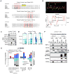
Figure 1. WASp nucleocytosolic transport sequences and partner proteins
(A) Putative NLS- and NES-motifs (underlined) of human WASp aligned with WASp-like proteins of other species. The evolutionary conserved core amino acids known to impart NLS- or NES-specific function are indicated in red. The “ϕ” in the NESs denote hydrophobic amino acid. (B) The hydropathy profiles of the putative NLS and NES-motifs of human WASp computed from the standard algorithms of Kyte-Doolittle (in yellow) and Hopp-Woods (in pink). PONDR plot of WASp, which predicts intrinsically-disordered regions (IDRs) in WASp is also shown. A score > 0.5 predicts disordered domains and <0.5 ordered domains. (C) Subcellular fractionation of T cell compartments. The purity of the indicated fractions (CP, CM, NP, NM) isolated from TH1-skewed human primary cells probed with the indicated antibodies. CP, cytoplasm depleted of total cellular membranes; CM, purified cellular membranes; NP, nucleoplasm depleted of total cellular membranes; NM, purified nuclear membranes. (D) Coumassie staining of proteins IP’ed with anti-WASp or control IgG antibody from the indicated fractions of primary TH1-skewed cells. A ~65kDa band corresponding to WASp is indicated by an asterisk. Excised bands were analyzed by MS. (E) The actual number of polypeptides of WASp-associated, nucleocytoplasmic transport proteins in the cell fractions are shown for cytosolic and nuclear WASp-complexes immunoprecipitated (IP) with anti-WASp, -FLAG/MYC (2-step immunoaffinity purification), or -IgG Abs. (F) Validation of MS-generated WASp-transport proteome by Co-IP. Protein complexes IP’ed by anti-WASp or control Ig antibody from TH1-skewed cells were resolved by sequential Western blotting the same gel using indicated antibodies. Loaded IP material is ~10% of input.
Figure 2
Figure 2. Functional validation of NLS- and NES-motifs
(A) Sequential Western blotting with the indicated antibodies on the nuclear (nu) and cytosolic (cyt) fractions generated from WAS TH cells expressing either full-length (FL) or the indicated Flag/Myc-tagged WASp-mutants (Δ) or untransfected (UT), activated under TH1-skewing or non-skewing TH0 conditions. (B) Cytosolic and nuclear fractions of Jurkat (TH1-skewed, TCR-activated) cells expressing Flag-tagged FL-WASp or its indicated WASp mutants were IP’ed with anti-Flag or -IgG Ab, and analyzed by sequential Western blotting with the indicated antibodies. Loaded IP is ~10% of input. The purity of the fractions was verified by Histone and LAMP-1 staining, which is shown in panel A. (C) Deconvolution fluorescence images of TH1-skewed (TCR-activated), human WASnull TH cells expressing the indicated WASp mutants. The images shown are after collapsing the entire z-stack images acquired at 0.2μm step size. Arrows point to cells displaying prominent nuclear WASp signal. Between 20–30 cells were analyzed. (D) Coimmunoprecipitation was performed with anti-FLAG or -IgG (control) antibodies from the cytosolic and nuclear fractions of Jurkat TH cells reconstituted with the indicated mutants and activated under TH1-skewing conditions. Serial immunoblotting was performed with the indicated antibodies. IP loading was 10% of the total input. The data is representative of at least 2 independent experiments. (E) MT/Dynein inhibition assays. Serial western blotting with the indicated antibodies of the nuclear (nu) and cytoplasmic (cyt) fractions derived from primary human TH1-skewed cells after treating with the indicated pharmacological agents or their controls. (F) Serial western blotting with the indicated antibodies of the nuclear (NF) and cytosolic (CF) fractions of human primary TH1-skewed cells treated with Leptomycin B (LMB) or control (ctrl)/DMSO for indicated durations. The data is representative of two experiments. (G) Western blot: the description is similar to that for panel B.
Figure 3
Figure 3. Western-based, histone H3 HMTase, assay
Histone H3 methyltransferase activity of WASp IP’ed with anti-Flag antibody from the nu/cyt fractions of HeLa cells stably transfected with FL-WASp or the indicated WASp-mutants. HeLa core histones and material IP’ed with anti-RBBP5 antibody are the positive controls, whereas Flag-IP in the mock transfected cells and IgG-IP are the negative controls. Reaction mixtures were immunoblotted with the indicated series of antibodies. The data is representative of at least two independent assays from two separate transfection events.
Figure 4
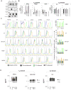
Figure 4. Characterizing the effect of WASp domain-deleted mutants on TH1- and TH2-activation
(A) Sequential western blotting with the indicated antibodies of the nuclear (nu) and cytosolic (cyt) fractions of human primary CD4+ TH cells, TH1-skewed, TH2-skewed, or non-skewed TH0 (all three CD3/28-activated). (B) RT-qPCR quantitation of candidate TH1- or TH2-genes in WASnull T cells reconstituted with FL-WASp or its indicated mutants after CD3/28-activation under TH1- or TH2-skewing or TH0 non-skewing conditions. Normal T cell line is the control. The mRNA copy numbers derived from the control TH0 cells are not shown, but were subtracted from the displayed final mRNA values of the TH1- or TH2-skewed cells. Absolute copy numbers adjusted to GAPDH are displayed as fold change (up or down) in TH1 or TH2 cells compared to their TH0 controls. Data represent the average of duplicates from at least 5 independent experiments from 3 separate transfections, with bars indicating SEM. Wilcoxon non-parametric test using the GraphPad InStat software determined the p-values comparing the data between WASnull T cells (UT) and FL/or mutant-expressing T cells (black asterisk, p<0.01) or between FL and mutants (red asterisk, p<0.01). In data where the differences did not reach statistical significance (i.e., p>0.05), asterisk is not shown (C) Flow cytometric histogram profiles showing expression of the indicated intracellular cytokines or transcription factors for TH0-nonskewed, TH1- and TH2-skewed, CD3/28-activated T cells transfected with the indicated WASp mutants. The bar graphs next to each histogram show the shift in mean fluorescence intensity (MFI) relative to their isotype controls and was quantified using the arithmetic average on a log scale (geometric mean). (D) Quantification of the secreted cytokines in the supernatants of cell cultures, whose mRNA profile is displayed in panel C, was assessed by quantitative ELISA performed in triplicates from at least 2 independent assays. Bars indicate SEM. Asterisk denotes p<0.05.
Figure 5
Figure 5. Chromatin-remodeling effects of WASp on its target gene loci
MNase-ChIP-qPCR. Chromatin enrichment profiles of the indicated proteins, at 5′UTR or 3′ exon ends of the indicated genes in TH1- or TH2-skewed cells, normal or WASnull TH cells, untransfected (UT) or stably transfected with the indicated WASp-mutants. The efficiency of MNase digested chromatin is displayed in Supplementary Fig. S2F. The displayed ChIP values (% of total input), shown as stacked columns, were derived after subtracting the background values obtained with isotype IgG antibody control ChIPs, the latter not shown. Data are expressed as percent immunoprecipitation relative to nuclear input chromatin (mean ± SEM) and represent an average of at least 5 independent experiments performed in duplicates from at least 3 separate transfection events. Intergenic region between COL8A2 and TRAPPC3 genes on Chr.1, which does not contain known protein-coding genes, served as a negative control. The genomic location of PCR primer/probes is indicated by red asterisk. For TBX21, the 5′ UTR primers were designed within the genomic region that also contains a GAS (γ-activated sequence) site (5′-TTCAGGCAA-3′ at about −770 bp from first coding ATG). For IFNG, the primers are located between −200 to −250 bp from first coding ATG, a region known to contain functional promoter elements (51). DNase I HS profile for the primary human peripheral TH1 cells (in grey) available from the ENCODE-University of Washington was aligned alongside our custom tracks to give context to the location of our ChIP-PCR primer/probes. Panel A: WASp and Transcription factors, Panel B: Histone modifications, and Panel C: RNA Pol II and SPT5. In panel C, 3′ denotes ChIP enrichment at the 3′ ends (last coding exon); 5′ denotes ChIP enrichment between 5′UTR and first coding exon.

Similar articles

Cited by

References

    1. Imai K, Morio T, Zhu Y, Jin Y, Itoh S, Kajiwara M, Yata J, Mizutani S, Ochs HD, Nonoyama S. Clinical course of patients with WASP gene mutations. Blood. 2004;103:456–464. - PubMed
    1. Catucci M, Castiello MC, Pala F, Bosticardo M, Villa A. Autoimmunity in wiskott-Aldrich syndrome: an unsolved enigma. Front Immunol. 2012;3:209. - PMC - PubMed
    1. Thrasher AJ, Burns SO. WASP: a key immunological multitasker. Nat Rev Immunol. 2010;10:182–192. - PubMed
    1. Higgs HN, Pollard TD. Regulation of actin filament network formation through ARP2/3 complex: activation by a diverse array of proteins. Annu Rev Biochem. 2001;70:649–676. - PubMed
    1. Campellone KG, Welch MD. A nucleator arms race: cellular control of actin assembly. Nat Rev Mol Cell Biol. 2010;11:237–251. - PMC - PubMed

Publication types

MeSH terms