. 2014 May 28;34(22):7485-92.
doi: 10.1523/JNEUROSCI.5294-13.2014.

Synaptic modifications in the medial prefrontal cortex in susceptibility and resilience to stress

Affiliations

Synaptic modifications in the medial prefrontal cortex in susceptibility and resilience to stress

Minghui Wang et al. J Neurosci. .

Abstract

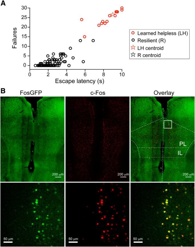
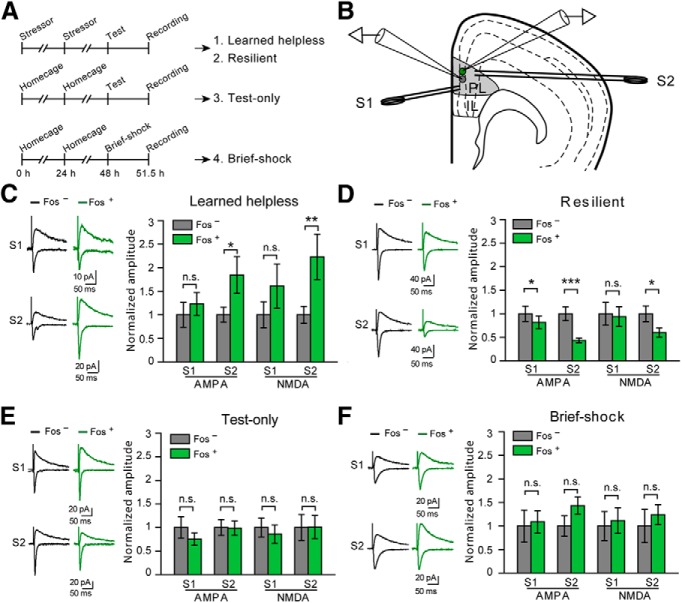
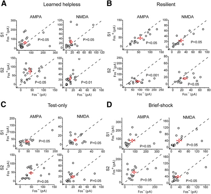
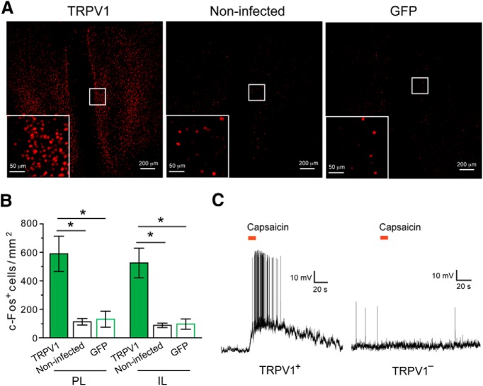
When facing stress, most individuals are resilient whereas others are prone to developing mood disorders. The brain mechanisms underlying such divergent behavioral responses remain unclear. Here we used the learned helplessness procedure in mice to examine the role of the medial prefrontal cortex (mPFC), a brain region highly implicated in both clinical and animal models of depression, in adaptive and maladaptive behavioral responses to stress. We found that uncontrollable and inescapable stress induced behavioral state-dependent changes in the excitatory synapses onto a subset of mPFC neurons: those that were activated during behavioral responses as indicated by their expression of the activity reporter c-Fos. Whereas synaptic potentiation was linked to learned helplessness, a depression-like behavior, synaptic weakening, was associated with resilience to stress. Notably, enhancing the activity of mPFC neurons using a chemical-genetic method was sufficient to convert the resilient behavior into helplessness. Our results provide direct evidence that mPFC dysfunction is linked to maladaptive behavioral responses to stress, and suggest that enhanced excitatory synaptic drive onto mPFC neurons may underlie the previously reported hyperactivity of this brain region in depression.

Keywords: c-Fos; chemical–genetic; depression; excitatory synapses; learned helplessness; mPFC.


Figures

Figure 1.
Figure 1.
The learned helplessness procedure induces behavioral changes and mPFC neuronal activation. A, A group of 110 FosGFP mice were subjected to the learned helplessness procedure, and their performance was analyzed by k-means (k = 2) clustering, using the number of failures and escape latency as classification parameters. Mice were classified as being either resilient (R) or learned helpless (LH) (see Materials and Methods). Among these 110 mice, 23 (red circles) belong to the LH group, and the rest (black circles) to the R group. Red and black stars represent the centroids for the learned helpless and the resilient group, respectively. B, Representative images of c-Fos expression in the mPFC of a FosGFP mouse killed 3.5 h after the learned helplessness testing session. Left, Expression of FosGFP was detected on the basis of GFP intrinsic fluorescence. Middle, c-Fos expression was detected using an antibody recognizing c-Fos. Right, Overlay, Bottom panels: higher-magnification images of the boxed area in PL (prelimbic cortex).
Figure 2.
Figure 2.
Synaptic modifications in the mPFC associated with susceptibility and resilience to learned helplessness. A, A schematic of the experimental procedure. Top, Mice were subjected to the learned helplessness procedure and were separated into “learned helpless” (1) and “resilient” (2) groups (see Materials and Methods). Middle, Mice were exposed only to the learned helplessness testing session (“test-only”) (3). Bottom, Mice were exposed to 10 brief foot shocks (“brief-shock”) (4). B, Schematic of the recording configuration. Synaptic responses onto a Fos+ (green) cell and that onto a Fos (gray) cell in the PL are simultaneously recorded. Synaptic transmission is evoked by two stimulating electrodes: one (S1) placed in layer 2/3 and the other (S2) in layer 5/6. C, Data from the “learned helpless” group. Left, Representative traces of EPSCs, which were recorded from a Fos+ neuron (green) and an adjacent Fos neuron (black), and which were evoked by S1 (top) and S2 (bottom). Right, Quantification of AMPAR- and NMDAR-mediated EPSC amplitude, which is normalized to the mean EPSC amplitude of Fos neurons. EPSCs are larger in Fos+ cells in response to S2 stimulation. D, Data from the “resilient” group. Data presentation is the same as that in C. EPSCs onto Fos+ cells were smaller than those onto Fos cells. E, Data from “test-only” group. Data presentation is the same as that in C. No significant difference was found. F, Data from “brief-shock” group. Data presentation is the same as that in C. No significant difference was found. *p < 0.05. **p < 0.01. ***p < 0.001. n.s., Nonsignificant.
Figure 3.
Figure 3.
mPFC synaptic modifications in susceptibility and resilience to learned helplessness. A–D, The same electrophysiology data as those in Figure 2C–F, respectively, are presented as scatter plots and in absolute values of EPSC amplitude. Each circle represents amplitudes of EPSCs recorded from a pair of Fos+/Fos neurons. A–D, Left, AMPAR-mediated EPSCs. Right, NMDAR-mediated EPSCs. Top, EPSCs evoked by S1. Bottom, EPSCs evoked by S2. Red circles represent mean ± SEM.
Figure 4.
Figure 4.
Chemical–genetic activation of mPFC neurons in ROSA-stopflox-TRPV1; TRPV1−/− mice converts resilience to helplessness-like behavior. A, Representative image of the mPFC of a ROSA-stopflox-TRPV1;TRPV1−/− mouse with bilateral injections of AAV-GFP-IRES-Cre to activate TRPV expression. B, Capsaicin treatment (20 mg/kg, i.p.) of the injected mice markedly increased both the number of failures and escape latency in the learned helplessness testing session. These behavioral parameters for each mouse are represented by a pair of circles, with the black and red circles indicating parameters measured in the absence and presence, respectively, of capsaicin treatment. Dashed line connects circles representing the same mouse. Compared with the black circles, the red circles are closer to the LH centroid (red star, reproduced from Fig. 1A for visual inspection). C, Behavioral effect of capsaicin treatment, quantified as the squared M-distance to the LH centroid (Fig. 1A; see Materials and Methods), was dose-dependent and reversible. The same mice were tested for learned helplessness at 5 min after each of the capsaicin treatment at the indicated dose, and also at 1 h for 20 mg/kg only. D, Chemical–genetic activation of mPFC neurons does not affect motor activity in an open field test. The “TRPV1” mice are the same ROSA-stopflox-TRPV1;TRPV1−/− mice (in which the mPFC were injected with AAV-GFP-IRES-Cre) used in B and C. The “Control” mice are ROSA-stopflox-TRPV1;TRPV1−/− mice without viral injection. At 5 min after treatment with capsaicin, mice were tested in an open field test. The total distance traveled (left), distance traveled in the center (middle), and velocity in the center (right) of the arena were measured. Activation of mPFC neurons by capsaicin did not significantly affect these parameters. E, Resilient mice were repeatedly tested in the learned helplessness testing sessions, with a 2 d intersession interval. Behavioral responses are quantified as in C. The resilient behavior did not change with repeated testing. ***p < 0.001.
Figure 5.
Figure 5.
Administration of capsaicin alone does not affect the behavior of the ROSA-stopflox-TRPV1;TRPV1−/− mice. A, Behavioral responses were quantified as in Figure 4C. Capsaicin treatment did not change the behavioral responses of ROSA-stopflox-TRPV1;TRPV1−/− mice that did not receive virus injection. Each circle represents one mouse; some of the circles are overlapping. B, Same as A, except that the ROSA-stopflox-TRPV1;TRPV1−/− mice were injected with an AAV-GFP into the mPFC. Some of the circles are overlapping.
Figure 6.
Figure 6.
TRPV1-mediated activation of mPFC neurons in the ROSA-stopflox-TRPV1;TRPV1−/− mice. A, Representative images of c-Fos expression in the mPFC after capsaicin treatment. Left, ROSA-stopflox-TRPV1;TRPV1−/− mice in which the mPFC was injected with AAV-GFP-IRES-Cre (to activate TRPV1 expression). Middle, ROSA-stopflox-TRPV1;TRPV1−/− mice that did not receive viral injection. Right, ROSA-stopflox-TRPV1;TRPV1−/− mice in which the mPFC was injected with AAV-GFP. Inset, Higher-magnification image of the boxed area. c-Fos was recognized by an antibody. B, Capsaicin treatment activated mPFC neurons as indicated by c-Fos expression in both PL and IL areas. C, The mPFC of ROSA-stopflox-TRPV1;TRPV1−/− mice was injected with AAV-GFP-IRES-Cre to induce TRPV1 expression. Puffs of capsaicin (8 μm, indicated by the red bars) applied to the cell body induced a robust increase in spiking activity in the majority of TRPV1-positive (TRPV1+) mPFC neurons (10 of 12 neurons from 6 mice) (left) but did not change activity in any of the TRPV1-negative (TRPV1) mPFC neurons recorded (13 of 13 neurons from 6 mice) (right). *p < 0.05.

References

    1. Amat J, Sparks PD, Matus-Amat P, Griggs J, Watkins LR, Maier SF. The role of the habenular complex in the elevation of dorsal raphe nucleus serotonin and the changes in the behavioral responses produced by uncontrollable stress. Brain Res. 2001;917:118–126. doi: 10.1016/S0006-8993(01)02934-1. - DOI - PubMed
    1. Amat J, Baratta MV, Paul E, Bland ST, Watkins LR, Maier SF. Medial prefrontal cortex determines how stressor controllability affects behavior and dorsal raphe nucleus. Nat Neurosci. 2005;8:365–371. doi: 10.1038/nn1399. - DOI - PubMed
    1. Arenkiel BR, Klein ME, Davison IG, Katz LC, Ehlers MD. Genetic control of neuronal activity in mice conditionally expressing TRPV1. Nat Methods. 2008;5:299–302. doi: 10.1038/nmeth.1190. - DOI - PMC - PubMed
    1. Barth AL. Visualizing circuits and systems using transgenic reporters of neural activity. Curr Opin Neurobiol. 2007;17:567–571. doi: 10.1016/j.conb.2007.10.003. - DOI - PMC - PubMed
    1. Birder LA, Nakamura Y, Kiss S, Nealen ML, Barrick S, Kanai AJ, Wang E, Ruiz G, De Groat WC, Apodaca G, Watkins S, Caterina MJ. Altered urinary bladder function in mice lacking the vanilloid receptor TRPV1. Nat Neurosci. 2002;5:856–860. doi: 10.1038/nn902. - DOI - PubMed

Publication types