. 2014 May 28;34(22):7663-76.
doi: 10.1523/JNEUROSCI.0718-14.2014.

Orbitofrontal dopamine depletion upregulates caudate dopamine and alters behavior via changes in reinforcement sensitivity

Affiliations

Orbitofrontal dopamine depletion upregulates caudate dopamine and alters behavior via changes in reinforcement sensitivity

H F Clarke et al. J Neurosci. .

Abstract

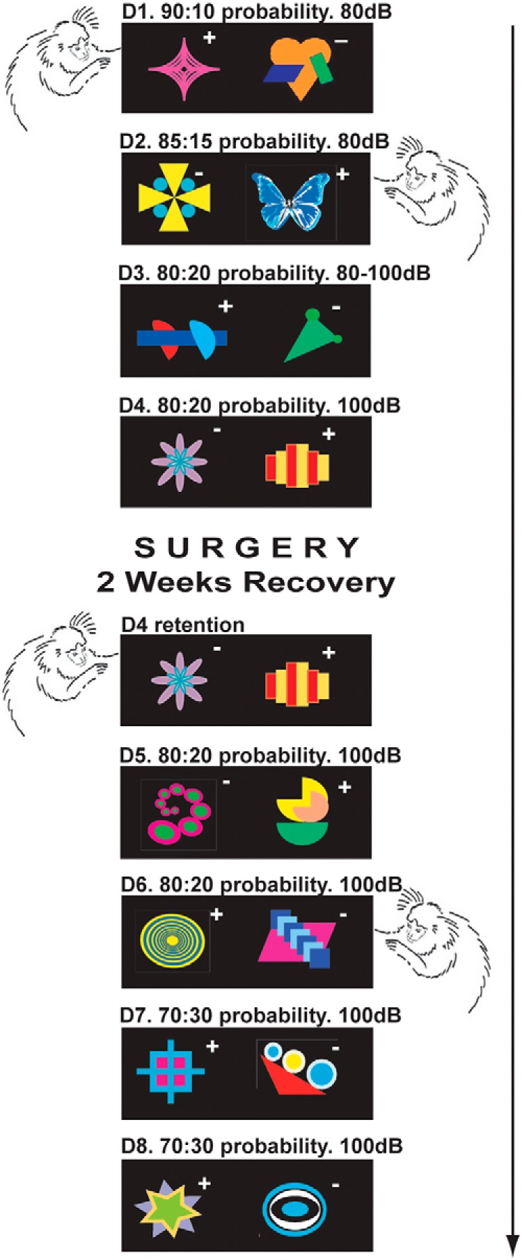
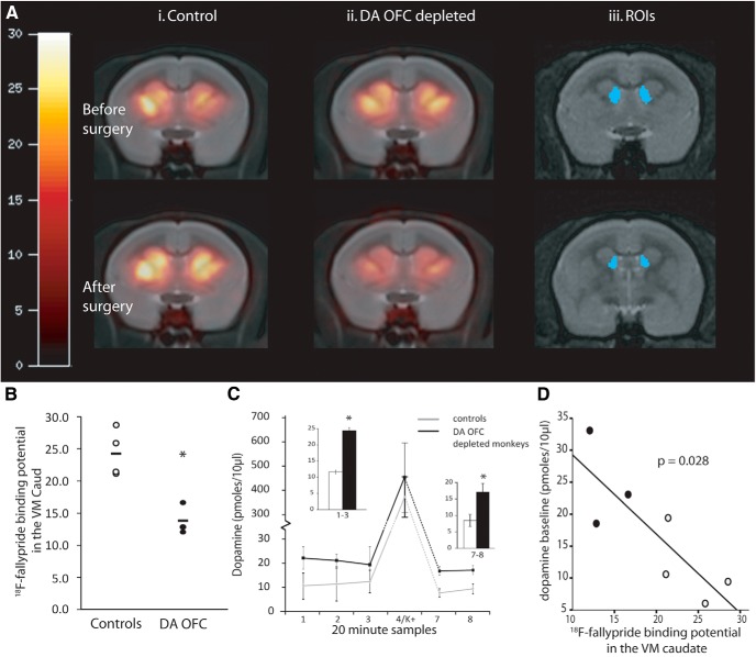
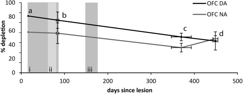
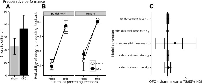
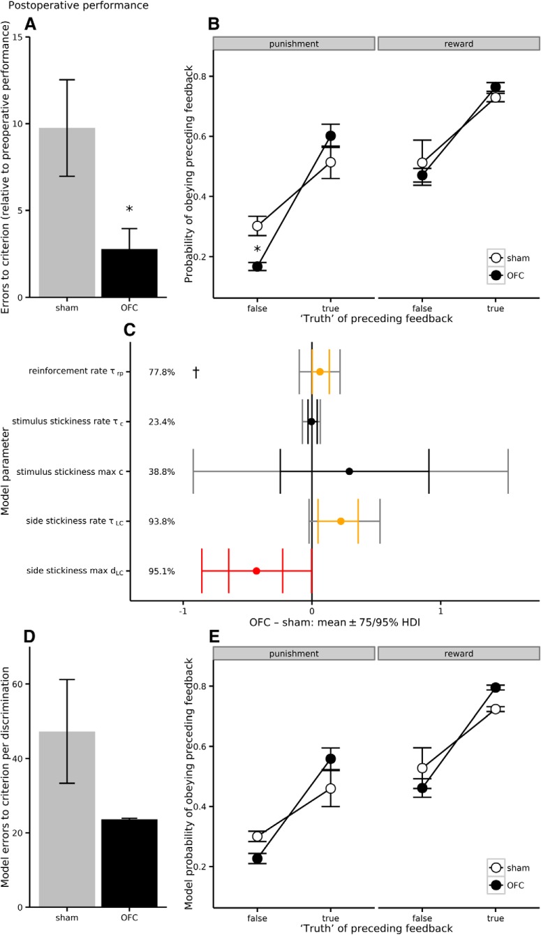
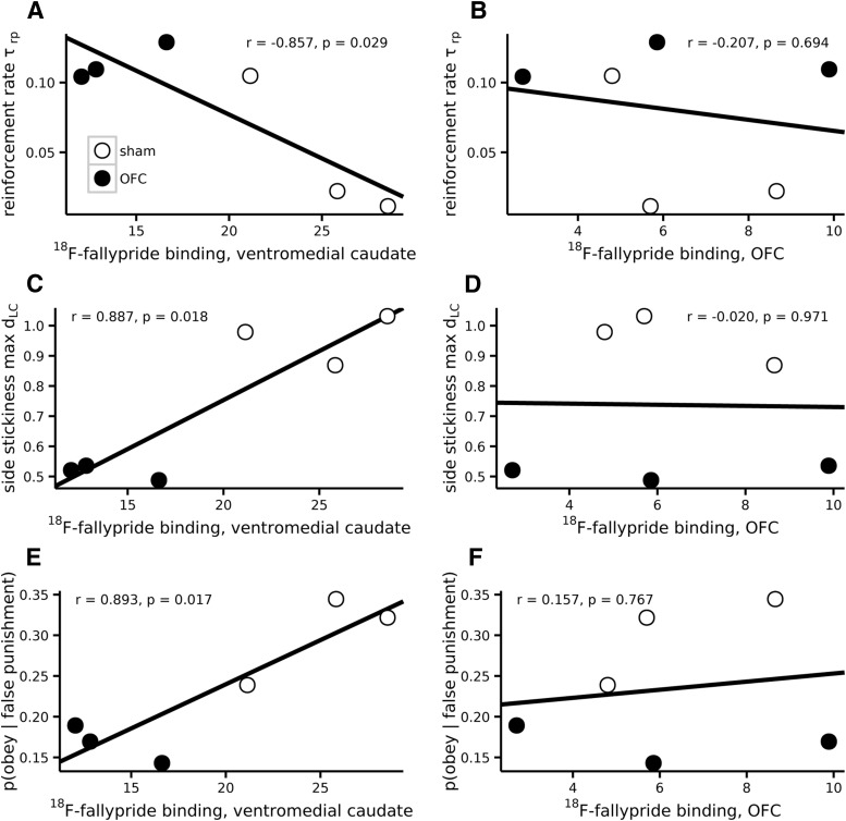
Schizophrenia is associated with upregulation of dopamine (DA) release in the caudate nucleus. The caudate has dense connections with the orbitofrontal cortex (OFC) via the frontostriatal loops, and both areas exhibit pathophysiological change in schizophrenia. Despite evidence that abnormalities in dopaminergic neurotransmission and prefrontal cortex function co-occur in schizophrenia, the influence of OFC DA on caudate DA and reinforcement processing is poorly understood. To test the hypothesis that OFC dopaminergic dysfunction disrupts caudate dopamine function, we selectively depleted dopamine from the OFC of marmoset monkeys and measured striatal extracellular dopamine levels (using microdialysis) and dopamine D2/D3 receptor binding (using positron emission tomography), while modeling reinforcement-related behavior in a discrimination learning paradigm. OFC dopamine depletion caused an increase in tonic dopamine levels in the caudate nucleus and a corresponding reduction in D2/D3 receptor binding. Computational modeling of behavior showed that the lesion increased response exploration, reducing the tendency to persist with a recently chosen response side. This effect is akin to increased response switching previously seen in schizophrenia and was correlated with striatal but not OFC D2/D3 receptor binding. These results demonstrate that OFC dopamine depletion is sufficient to induce striatal hyperdopaminergia and changes in reinforcement learning relevant to schizophrenia.

Keywords: PET; behavior; caudate nucleus; dopamine; orbitofrontal cortex; schizophrenia.


Figures

Figure 1.
Figure 1.
Task sequence (D, discrimination). Representative stimuli are shown, labeled + for correct and − for incorrect. Reinforcement probabilities are shown: for example, “90:10 probability” indicates that P(reward | correct stimulus selected) = P(punishment | incorrect stimulus selected) = 0.9 and P(punishment | correct stimulus selected) = P(reward | incorrect stimulus selected) = 0.1. The intensity of auditory punishment is shown in dB SPL.
Figure 2.
Figure 2.
Increased ventromedial caudate dopamine levels following 6-OHDA lesions of the OFC, shown by reduced binding potential of the selective D2/3 receptor antagonist 18F-fallypride and increased baseline extracellular DA levels. A, Coronal coregistered MRI and PET images of 18F-fallypride nondisplaceable binding potential (BPND) in the striatum of a control animal (Ai), and an OFC DA-depleted animal (Aii), before and after surgery, depicted at coronal section AP +10. Aiii, Ventromedial caudate ROIs (blue) are depicted at coronal section AP +10 (top) and AP +9.75 (bottom); BPND threshold = 30. B, Significantly reduced 18F-fallypride BPND in the ventromedial caudate of OFC DA-depleted monkeys, compared with controls (*p < 0.05). C, Significantly increased baseline extracellular DA in the ventromedial (VM) caudate both before and after the K+ challenge of OFC DA-depleted monkeys, measured by microdialysis (insets: samples 1–3, p = 0.041; samples 7–8, p = 0.04). D, Extracellular DA levels before K+ challenge correlated negatively with 18F-fallypride BPND in the ventromedial caudate (p = 0.028; filled circles, OFC DA-depleted group; open circles, controls).
Figure 3.
Figure 3.
Postmortem depletions of DA and NA in the OFC as a function of time since surgery in OFC-depleted monkeys. The gray regions indicate the time periods in which the behavior (i), the second MRI/PET scan (ii), and in vivo microdialysis (iii) were completed by the DA OFC depleted monkeys. Each gray region extends from the earliest starting point to the latest endpoint (and for behavior, their edges represent the monkeys that were the “fastest” and “slowest” to complete the discriminations). All three components of this study therefore occurred during high levels of OFC DA depletion. a, One DA OFC-depleted animal, 16 d after surgery; b, four DA OFC-depleted animals averaging 84 d after surgery (Clarke et al., 2007); c, Four DA OFC-depleted animals averaging 370 d after surgery (Walker et al., 2009); and d, current behavioral study.
Figure 4.
Figure 4.
Preoperative behavioral performance. The monkeys did not show any group differences (p > 0.05) in either (A) their ability to learn the preoperative probabilistic discriminations (D1–D4; compare Fig. 5A), or (B) their win-stay/lose-shift behavior (last preoperative discrimination, D4; compare Fig. 5B). C, Similarly, there were no group differences in parameters for the best computational model when fitted to preoperative discrimination D4 except a fractionally higher τc (75% HDI [0.0025, 0.051], 95% HDI [−0.019, 0.081]; HDIs for all other parameters included zero; compare Fig. 5C).
Figure 5.
Figure 5.
Behavioral performance. A, Faster learning in OFC DA-depleted monkeys in a probabilistic visual discrimination learning task, with fewer errors to criterion compared with their preoperative performance (p ≤ 0.05). B, OFC DA-depleted monkeys showed intact reward-related behavior but a decreased probability of shifting their responding to the other stimulus after misleading (false) negative feedback (†p = 0.018). C, The optimal computational model of behavior had parameters representing sensitivity to reinforcement (τrp), a tendency to repeat choices to recently chosen stimuli (τc, c), and a tendency to repeat choices to recently chosen sides (τLC, dLC). Lesioned subjects showed increased sensitivity to reinforcement (higher τrp). They also showed less side stickiness (shown both by a lower dLC, indicating a reduction in the overall influence of side stickiness compared with that of reinforcement, and a higher τLC, indicating that the influence of side stickiness was less long-lasting). The dagger (†) indicates that between-group differences in τrp were necessary and sufficient for the other behavioral effects shown in D, E (see Materials and Methods, and Results). Error bars show the posterior distributions of group differences in group mean parameter values, as highest-density intervals (HDIs; orange, 75% HDI excludes zero; red, 95% HDI excludes zero). Percentages are the posterior probabilities that the parameter differs from zero (width of the largest HDI excluding zero), as described in the Materials and Methods; they are not frequentist p values. D, This computational model predicted fewer errors to criterion in the OFC DA-depleted group (compare with A). E, Moreover, the computational model predicted the differences in responding to false punishment in the behavioral data (compare with B).
Figure 6.
Figure 6.
Relationship between behavior and striatal dopamine. A, The dLC parameter correlated with 18F-fallypride BPND in the caudate (ventromedial caudate shown) but (B) not the OFC. C, Similarly, the τrp parameter correlated with 18F-fallypride BPND in the caudate but (D) not the OFC. The probability of shifting after false-negative feedback correlated with (E) the reduced levels of ventromedial caudate 18F-fallypride BPND seen in the OFC DA-depleted monkeys but not (F) the levels of 18F-fallypride BPND seen in the OFC.

Similar articles

Cited by

References

    1. Bertolino A, Knable MB, Saunders RC, Callicott JH, Kolachana B, Mattay VS, Bachevalier J, Frank JA, Egan M, Weinberger DR. The relationship between dorsolateral prefrontal N-acetylaspartate measures and striatal dopamine activity in schizophrenia. Biol Psychiatry. 1999;45:660–667. doi: 10.1016/S0006-3223(98)00380-1. - DOI - PubMed
    1. Bertolino A, Roffman JL, Lipska BK, van Gelderen P, Olson A, Weinberger DR. Reduced N-acetylaspartate in prefrontal cortex of adult rats with neonatal hippocampal damage. Cereb Cortex. 2002;12:983–990. doi: 10.1093/cercor/12.9.983. - DOI - PubMed
    1. Bouthenet ML, Souil E, Martres MP, Sokoloff P, Giros B, Schwartz JC. Localization of dopamine D3 receptor mRNA in the rat brain using in situ hybridization histochemistry: comparison with dopamine D2 receptor mRNA. Brain Res. 1991;564:203–219. doi: 10.1016/0006-8993(91)91456-B. - DOI - PubMed
    1. Burnham KP, Anderson DR. Multimodel inference: understanding AIC and BIC in model selection. Sociol Methods Res. 2004;33:261–304. doi: 10.1177/0049124104268644. - DOI
    1. Cardinal RN, Parkinson JA, Hall J, Everitt BJ. Emotion and motivation: the role of the amygdala, ventral striatum, and prefrontal cortex. Neurosci Biobehav Rev. 2002;26:321–352. doi: 10.1016/S0149-7634(02)00007-6. - DOI - PubMed

Publication types

LinkOut - more resources